WB
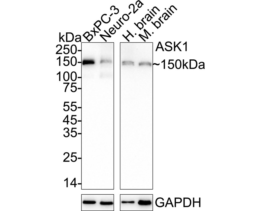
Western blot analysis of ASK1 on different lysates with Rabbit anti-ASK1 antibody at 1/1,000 dilution. Lane 1: BxPC-3 cell lysate, Lane 2: Neuro-2a cell lysate, Lane 3: Human brain tissue lysate, Lane 4: Mouse brain tissue lysate, Lysates/proteins at 20 µg/Lane. Exposure time: 3 minutes; 4-20% SDS-PAGE gel. Proteins were transferred to a PVDF membrane and blocked with 5% NFDM/TBST for 1 hour at room temperature. The primary antibody at 1/1,000 dilution was used in 5% NFDM/TBST at 4℃ overnight. Goat Anti-Rabbit IgG - HRP Secondary Antibody at 1/50,000 dilution was used for 1 hour at room temperature.IHC
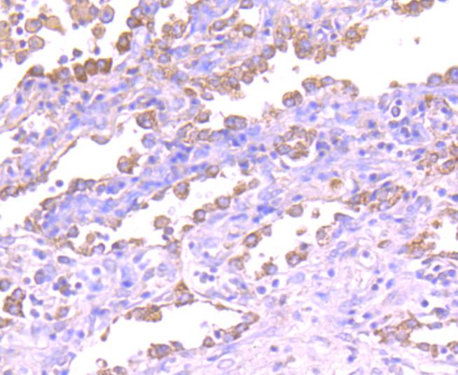
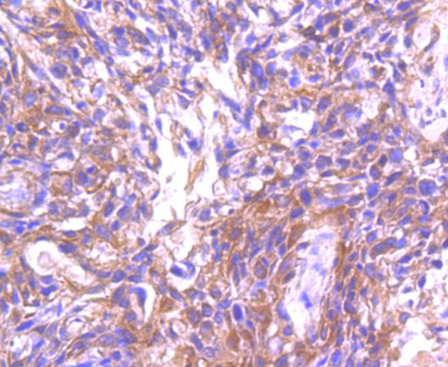
Immunohistochemical analysis of paraffin-embedded human lung cancer tissue using anti-ASK1 antibody. Counter stained with hematoxylin.IHC
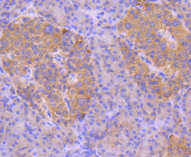
Immunohistochemical analysis of paraffin-embedded human pancreas tissue using anti-ASK1 antibody. Counter stained with hematoxylin.IHC
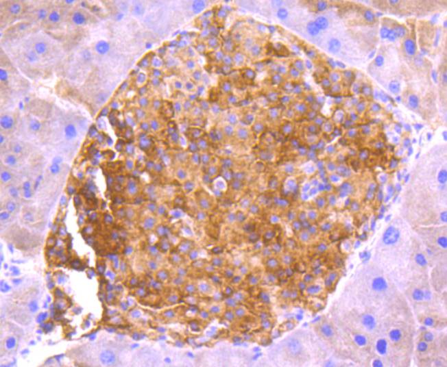
Immunohistochemical analysis of paraffin-embedded mouse pancreas tissue using anti-ASK1 antibody. Counter stained with hematoxylin.IHC
Immunohistochemical analysis of paraffin-embedded human breast carcinoma tissue using anti-ASK1 antibody. Counter stained with hematoxylin.| Product Name | ASK1 Recombinant Rabbit Monoclonal Antibody |
|---|---|
| Antibody Type | Primary Antibodies |
| Product description | Mitogen-activated protein (MAP) kinase cascades are activated by various extracellular stimuli including growth factors. The MEK kinases (also designated MAP kinase kinase kinases, MKKKs, MAP3Ks or MEKKs) phosphorylate and thereby activate the MEKs (also called MAP kinase kinases or MKKs), including ERK, JNK and p38. These activated MEKs in turn phosphorylate and activate the MAP kinases. The MEK kinases include Raf-1, Raf-B, Mos, MEK kinase-1, MEK kinase-2, MEK kinase-3, MEK kinase-4, ASK 1 (MEK kinase-5) and MAP3K6 (MEK kinase-6). MEK kinase-1 has been shown to phosphorylate MEK-1 via a Raf-independent pathway. Evidence suggests that MEK-3 is preferentially activated by MEK kinase-3 and that MEK-4 is activated by both MEK kinase-2 and MEK kinase-3. MEK kinase-4 has been shown to specifically activate the JNK pathway. ASK1 activates both MEK-4 and MEK-3/MEK-6 pathways. |
| Immunogen | Synthetic peptide within N-terminal human ASK1. |
| Clonality | monoclonal |
|---|---|
| Isotype | IgG |
| Host Species | Recombinant rabbit |
| Tested Applications | IHCWB |
| WB:1:500-1:2000 IHC:1:50-1:200 |
|
| Species Reactivity | HumanMouse |
| Concentration | 1mg/ml |
| Purification | Protein A |
| Alternative Names | Apoptosis signal regulating kinase 1 antibody Apoptosis signal-regulating kinase 1 antibody ASK 1 antibody ASK-1 antibody ASK1 antibody M3K5 antibody M3K5_HUMAN antibody MAP/ERK kinase kinase 5 antibody MAP3K5 antibody MAPK/ERK kinase kinase 5 antibody MAPKKK5 antibody MEK kinase 5 antibody MEKK 5 antibody MEKK5 antibody Mitogen activated protein kinase kinase kinase 5 antibody Mitogen-activated protein kinase kinase kinase 5 antibody |
|---|---|
| Molecular Weight(MW) | 155kDa(Observed band size: 150kDa) |
| Cellular Localization | Endoplasmic reticulum, Cytoplasm. |
WB
Western blot analysis of ASK1 on different lysates with Rabbit anti-ASK1 antibody at 1/1,000 dilution. Lane 1: BxPC-3 cell lysate, Lane 2: Neuro-2a cell lysate, Lane 3: Human brain tissue lysate, Lane 4: Mouse brain tissue lysate, Lysates/proteins at 20 µg/Lane. Exposure time: 3 minutes; 4-20% SDS-PAGE gel. Proteins were transferred to a PVDF membrane and blocked with 5% NFDM/TBST for 1 hour at room temperature. The primary antibody at 1/1,000 dilution was used in 5% NFDM/TBST at 4℃ overnight. Goat Anti-Rabbit IgG - HRP Secondary Antibody at 1/50,000 dilution was used for 1 hour at room temperature.
IHC
Immunohistochemical analysis of paraffin-embedded human lung cancer tissue using anti-ASK1 antibody. Counter stained with hematoxylin.
IHC
Immunohistochemical analysis of paraffin-embedded human pancreas tissue using anti-ASK1 antibody. Counter stained with hematoxylin.
IHC
Immunohistochemical analysis of paraffin-embedded mouse pancreas tissue using anti-ASK1 antibody. Counter stained with hematoxylin.
IHC
Immunohistochemical analysis of paraffin-embedded human breast carcinoma tissue using anti-ASK1 antibody. Counter stained with hematoxylin.| Application Notes | WB:1:500-1:2000 IHC:1:50-1:200 |
|---|
| Form | Liquid |
|---|---|
| Storage Instructions | Store at +4℃ after thawing. Aliquot store at -20℃ or -80℃. Avoid repeated freeze / thaw cycles. |
| Storage Buffer | 1*TBS (pH7.4), 1%BSA, 40%Glycerol. Preservative: 0.05% Sodium Azide. |


