Application
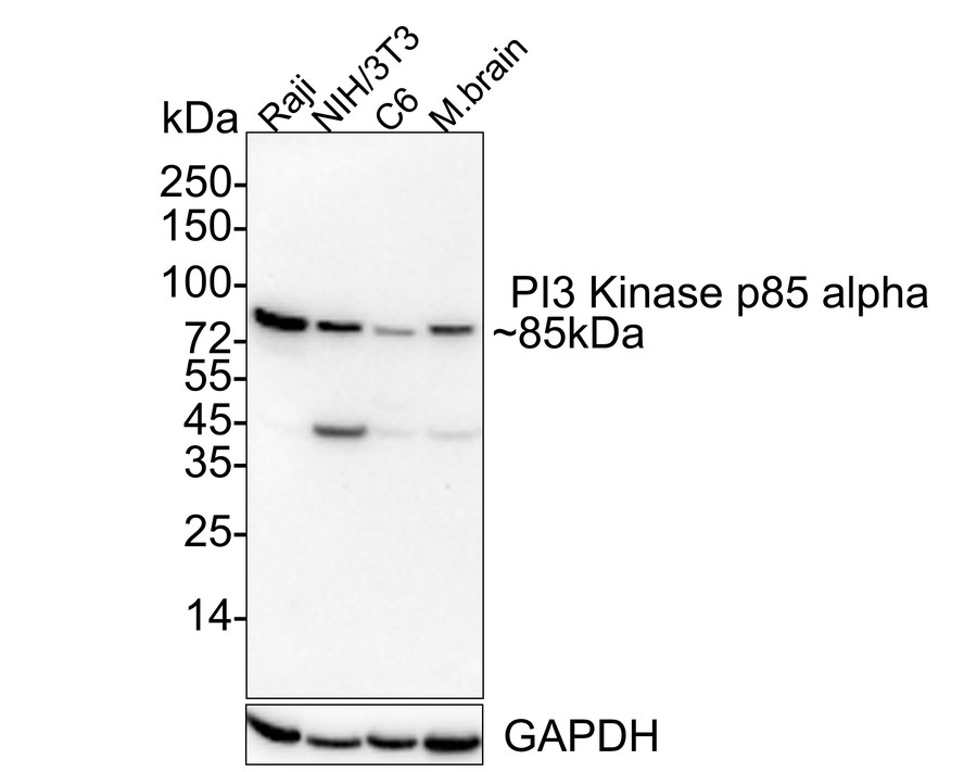
Fig1: Western blot analysis of PI 3 Kinase p85 alpha on different lysates with Rabbit anti-PI 3 Kinase p85 alpha antibody at 1/1,000 dilution. Lane 1: Raji cell lysate, Lane 2: NIH/3T3 cell lysate, Lane 3: C6 cell lysate, Lane 4: Mouse brain tissue lysate, Lysates/proteins at 20(cell lysate),40(tissue lysate) µg/Lane. Exposure time: 2 minutes; 4-20% SDS-PAGE gel. Proteins were transferred to a PVDF membrane and blocked with 5% NFDM/TBST for 1 hour at room temperature. The primary antibody at 1/1,000 dilution was used in 5% NFDM/TBST at 4℃ overnight. Goat Anti-Rabbit IgG - HRP Secondary Antibody at 1/50,000 dilution was used for 1 hour at room temperature.Application
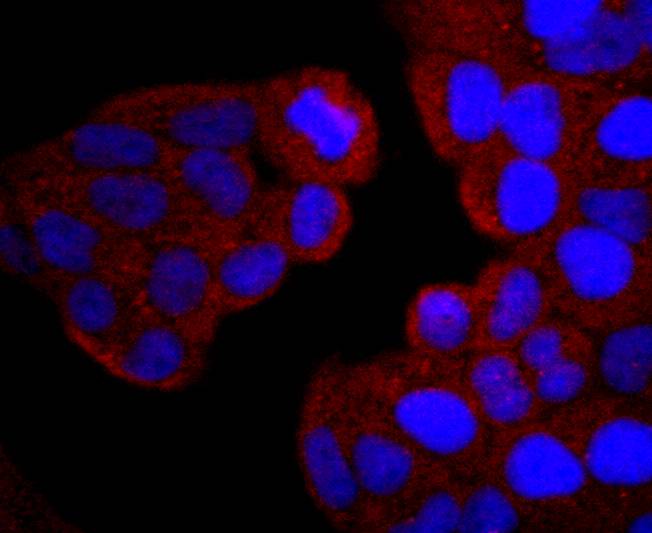
Fig2: ICC staining PI 3 Kinase p85 alpha in Hela cells (red). The nuclear counter stain is DAPI (blue). Cells were fixed in paraformaldehyde, permeabilised with 0.25% Triton X100/PBS.Application
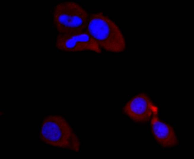
Fig3: ICC staining PI 3 Kinase p85 alpha in MCF-7 cells (red). The nuclear counter stain is DAPI (blue). Cells were fixed in paraformaldehyde, permeabilised with 0.25% Triton X100/PBS.Application
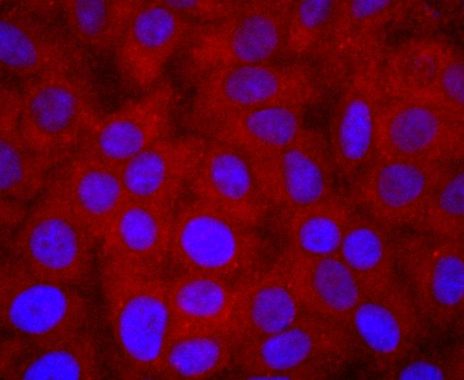
Fig4: ICC staining PI 3 Kinase p85 alpha in HepG2 cells (red). The nuclear counter stain is DAPI (blue). Cells were fixed in paraformaldehyde, permeabilised with 0.25% Triton X100/PBS.Application
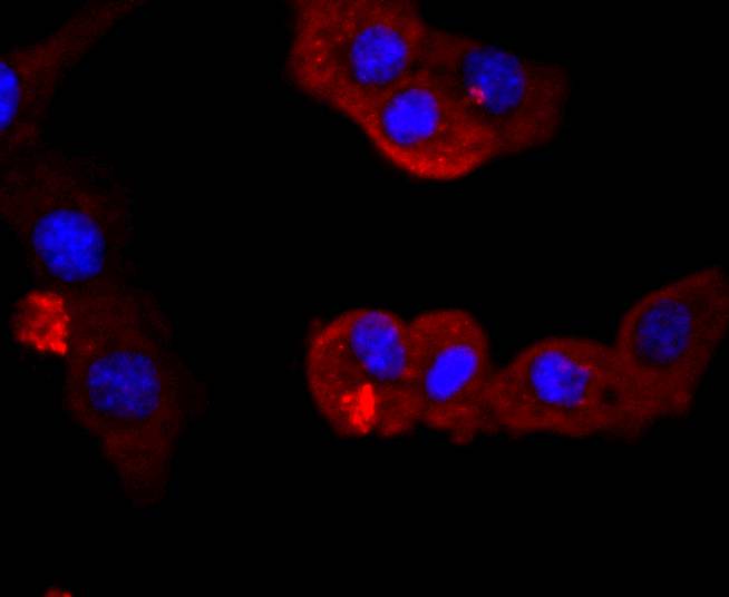
Fig5: ICC staining PI 3 Kinase p85 alpha in NIH/3T3 cells (red). The nuclear counter stain is DAPI (blue). Cells were fixed in paraformaldehyde, permeabilised with 0.25% Triton X100/PBS.Application
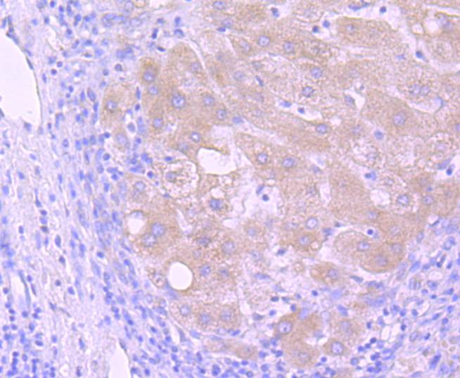
Fig6: Immunohistochemical analysis of paraffin-embedded human liver cancer tissue using anti-PI 3 Kinase p85 alpha antibody. Counter stained with hematoxylin.Application
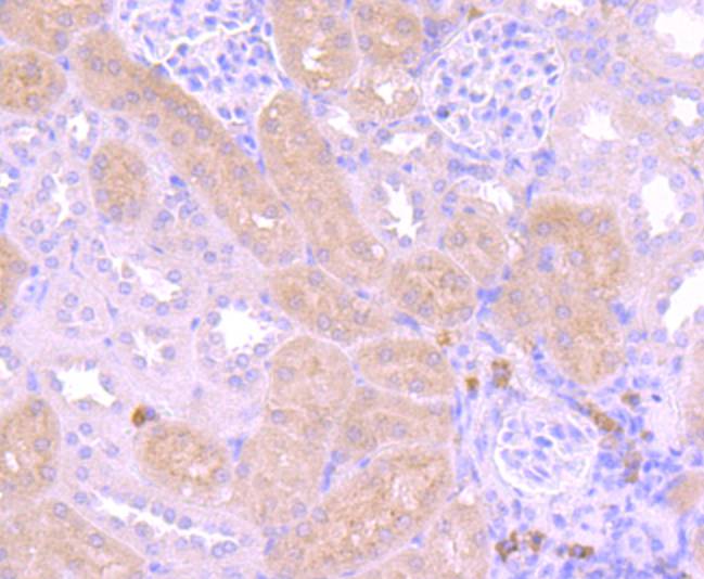
Fig7: Immunohistochemical analysis of paraffin-embedded human kidney tissue using anti-PI 3 Kinase p85 alpha antibody. Counter stained with hematoxylin.Application
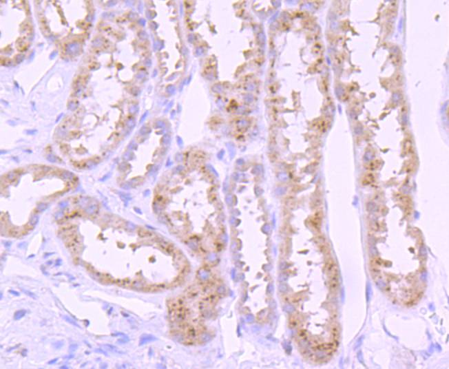
Fig8: Immunohistochemical analysis of paraffin-embedded mouse kidney tissue using anti-PI 3 Kinase p85 alpha antibody. Counter stained with hematoxylin.Application
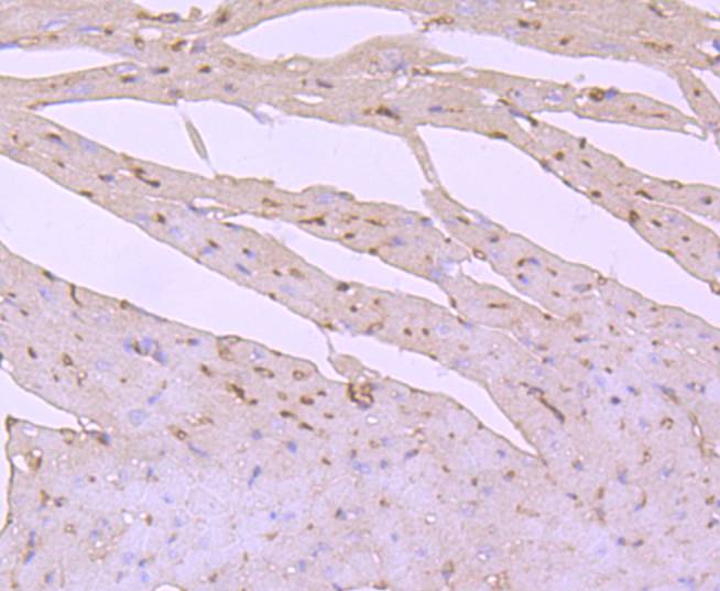
Fig9: Immunohistochemical analysis of paraffin-embedded mouse heart tissue using anti-PI 3 Kinase p85 alpha antibody. Counter stained with hematoxylin.Application
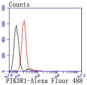
Fig10: Flow cytometric analysis of HepG2 cells with PI 3 Kinase p85 alpha antibody at 1/50 dilution (red) compared with an unlabelled control (cells without incubation with primary antibody; black). Alexa Fluor 488-conjugated goat anti rabbit IgG was used as the secondary antibody| Product Name | PI 3 Kinase p85 alpha Recombinant Rabbit Monoclonal Antibody |
|---|---|
| Antibody Type | Primary Antibodies |
| Product description | Phosphatidylinositol 3-kinase (PI 3-kinase) phosphorylates the 3' OH position of the inositol ring of inositol lipids and is composed of p85 and p110 subunits. PI 3-kinase p85 lacks PI 3-kinase activity and acts as an adapter, coupling p110 to activated protein tyrosine kinase. Two forms of p85 have been described (p85α and p85β), each possessing one SH3 and two SH2 domains. PI 3-kinase p85α, also known as GRB1, phosphatidylinositol 3-kinase regulatory 1 or p85, is a 724 amino acid protein that exists as four alternatively spliced isoforms. Involved in insulin metabolism, defects in the PI 3-kinase p85α gene have been linked to insulin resistance. PI 3-kinase p85α is polyubiquitinated in T-cells by Cbl-b, and has multiple phosphorylated amino acid residues, including a phosphorylated tyrosine residue at position 467. |
| Immunogen | Synthetic peptide within C-terminal human PI 3 Kinase p85 alpha. |
| Clonality | monoclonal |
|---|---|
| Isotype | IgG |
| Host Species | Recombinant rabbit |
| Tested Applications | FCICC/IFIHCWB |
| WB:1:1,000-1:2,000 ICC:1:50-1:200 IHC:1:50-1:200 FC:1:50-1:100 |
|
| Species Reactivity | HumanMouseRat |
| Concentration | 1mg/ml |
| Purification | Protein A |
| Alternative Names | GRB1 antibody
p85 alpha antibody
p85 antibody
P85A_HUMAN antibody
Phosphatidylinositol 3 kinase associated p 85 alpha antibody
Phosphatidylinositol 3 kinase regulatory 1 antibody
Phosphatidylinositol 3 kinase regulatory subunit polypeptide 1 (p85 alpha) antibody Phosphatidylinositol 3-kinase 85 kDa regulatory subunit alpha antibody Phosphatidylinositol 3-kinase regulatory subunit alpha antibody Phosphoinositide 3 kinase regulatory subunit 1 (alpha) antibody PI3 kinase p85 subunit alpha antibody PI3-kinase regulatory subunit alpha antibody PI3-kinase subunit p85-alpha antibody PI3K antibody PI3K regulatory subunit alpha antibody Pik3r1 antibody PtdIns 3 kinase p85 alpha antibody PtdIns-3-kinase regulatory subunit alpha antibody PtdIns-3-kinase regulatory subunit p85-alpha antibody |
|---|---|
| Molecular Weight(MW) | 84 kDa |
| Cellular Localization | Cytoplasm, membrane, nucleus. |
Application
Fig1: Western blot analysis of PI 3 Kinase p85 alpha on different lysates with Rabbit anti-PI 3 Kinase p85 alpha antibody at 1/1,000 dilution. Lane 1: Raji cell lysate, Lane 2: NIH/3T3 cell lysate, Lane 3: C6 cell lysate, Lane 4: Mouse brain tissue lysate, Lysates/proteins at 20(cell lysate),40(tissue lysate) µg/Lane. Exposure time: 2 minutes; 4-20% SDS-PAGE gel. Proteins were transferred to a PVDF membrane and blocked with 5% NFDM/TBST for 1 hour at room temperature. The primary antibody at 1/1,000 dilution was used in 5% NFDM/TBST at 4℃ overnight. Goat Anti-Rabbit IgG - HRP Secondary Antibody at 1/50,000 dilution was used for 1 hour at room temperature.
Application
Fig2: ICC staining PI 3 Kinase p85 alpha in Hela cells (red). The nuclear counter stain is DAPI (blue). Cells were fixed in paraformaldehyde, permeabilised with 0.25% Triton X100/PBS.
Application
Fig3: ICC staining PI 3 Kinase p85 alpha in MCF-7 cells (red). The nuclear counter stain is DAPI (blue). Cells were fixed in paraformaldehyde, permeabilised with 0.25% Triton X100/PBS.
Application
Fig4: ICC staining PI 3 Kinase p85 alpha in HepG2 cells (red). The nuclear counter stain is DAPI (blue). Cells were fixed in paraformaldehyde, permeabilised with 0.25% Triton X100/PBS.
Application
Fig5: ICC staining PI 3 Kinase p85 alpha in NIH/3T3 cells (red). The nuclear counter stain is DAPI (blue). Cells were fixed in paraformaldehyde, permeabilised with 0.25% Triton X100/PBS.
Application
Fig6: Immunohistochemical analysis of paraffin-embedded human liver cancer tissue using anti-PI 3 Kinase p85 alpha antibody. Counter stained with hematoxylin.
Application
Fig7: Immunohistochemical analysis of paraffin-embedded human kidney tissue using anti-PI 3 Kinase p85 alpha antibody. Counter stained with hematoxylin.
Application
Fig8: Immunohistochemical analysis of paraffin-embedded mouse kidney tissue using anti-PI 3 Kinase p85 alpha antibody. Counter stained with hematoxylin.
Application
Fig9: Immunohistochemical analysis of paraffin-embedded mouse heart tissue using anti-PI 3 Kinase p85 alpha antibody. Counter stained with hematoxylin.
Application
Fig10: Flow cytometric analysis of HepG2 cells with PI 3 Kinase p85 alpha antibody at 1/50 dilution (red) compared with an unlabelled control (cells without incubation with primary antibody; black). Alexa Fluor 488-conjugated goat anti rabbit IgG was used as the secondary antibody| Positive Control | Raji, MCF-7, Hela, MCF-7, HepG2, NIH/3T3, mouse kidney tissue, mouse heart tissue, human liver cancer tissue, human kidney tissue. |
|---|---|
| Application Notes | WB:1:1,000-1:2,000 ICC:1:50-1:200 IHC:1:50-1:200 FC:1:50-1:100 |
| Form | Liquid |
|---|---|
| Storage Instructions | Store at +4℃ after thawing. Aliquot store at -20℃ or -80℃. Avoid repeated freeze / thaw cycles. |
| Storage Buffer | 1*TBS (pH7.4), 1%BSA, 40%Glycerol. Preservative: 0.05% Sodium Azide. |


