WB
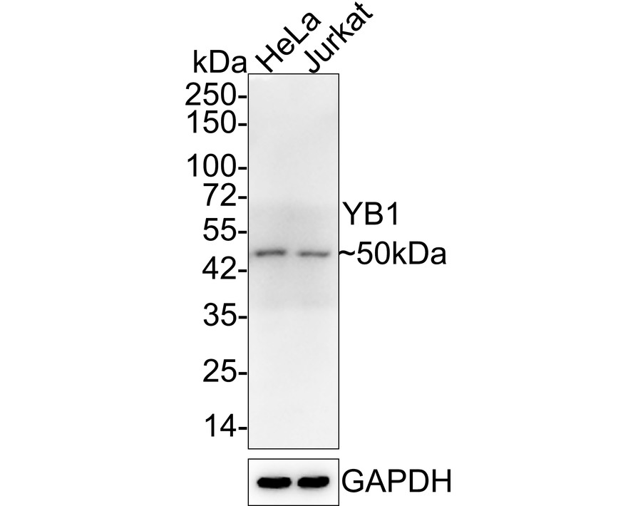
Western blot analysis of YB1 on different lysates with Rabbit anti-YB1 antibody at 1/1,000 dilution. Lane 1: HeLa cell lysate, Lane 2: Jurkat cell lysate, Lysates/proteins at 10 µg/Lane. Exposure time: 2 minutes 18 seconds; 4-20% SDS-PAGE gel. Proteins were transferred to a PVDF membrane and blocked with 5% NFDM/TBST for 1 hour at room temperature. The primary antibody at 1/1,000 dilution was used in 5% NFDM/TBST at 4℃ overnight. Goat Anti-Rabbit IgG - HRP Secondary Antibody at 1/50,000 dilution was used for 1 hour at room temperature.IHC
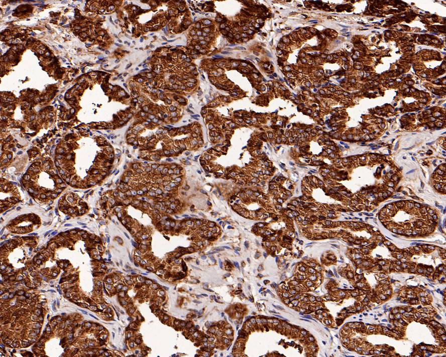
Immunohistochemical analysis of paraffin-embedded human prostate carcinoma tissue with Rabbit anti-YB1 antibody at 1/200 dilution. The section was pre-treated using heat mediated antigen retrieval with Tris-EDTA buffer (pH 9.0) for 20 minutes. The tissues were blocked in 1% BSA for 20 minutes at room temperature, washed with ddH2O and PBS, and then probed with the primary antibody at 1/200 dilution for 1 hour at room temperature. The detection was performed using an HRP conjugated compact polymer system. DAB was used as the chromogen. Tissues were counterstained with hematoxylin and mounted with DPX.IHC
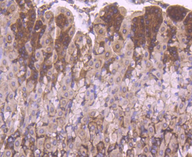
Immunohistochemical analysis of paraffin-embedded mouse stomach tissue using anti-YB1 antibody at 1/200. Counter stained with hematoxylin.ICC/IF
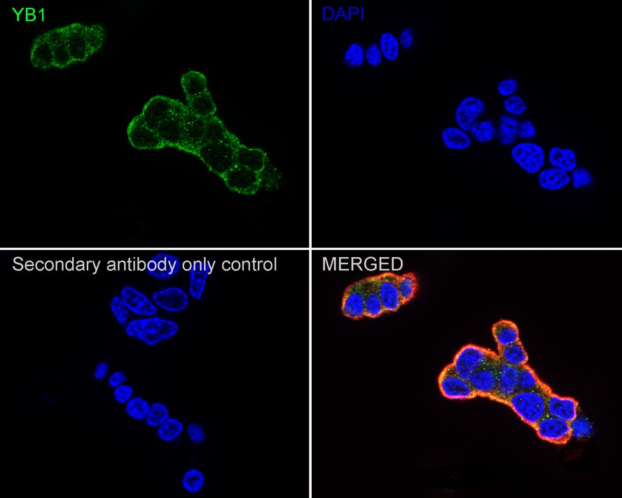
Immunocytochemistry analysis of PC-12 cells labeling YB1 with Rabbit anti-YB1 antibody at 1/100 dilution. Cells were fixed in 4% paraformaldehyde for 15 minutes at room temperature, permeabilized with 0.1% Triton X-100 in PBS for 15 minutes at room temperature, then blocked with 1% BSA in 10% negative goat serum for 1 hour at room temperature. Cells were then incubated with Rabbit anti-YB1 antibody at 1/100 dilution in 1% BSA in PBST overnight at 4 ℃. Goat Anti-Rabbit IgG H&L (488) was used as the secondary antibody at 1/1,000 dilution. PBS instead of the primary antibody was used as the secondary antibody only control. Nuclear DNA was labelled in blue with DAPI. Beta tubulin (red) was stained at 1/100 dilution overnight at +4℃. Goat Anti-Mouse IgG H&L (594) was used as the secondary antibody at 1/1,000 dilution.| Product Name | YB1 Recombinant Rabbit Monoclonal Antibody |
|---|---|
| Antibody Type | Primary Antibodies |
| Product description | Y-box binding protein YB-1 (also known as CCAAT-binding transcription factor, enhancer factor I subunit A and DNA-binding protein B) belongs to a family of multifunctional proteins which regulate both transcription and translation. Y-box proteins interact with a wide variety of nucleic acid structures to act as transcription factors and mRNA masking proteins. The modular structure of Y-box proteins includes a highly conserved N-terminal cold-shock domain (CSD, equivalent to the bacterial cold-shock proteins) and four basic C-terminal domains containing Arginine clusters and aromatic residues. YB-1 plays a role in cell proliferation as an activator of growth-associated gene expression. YB-1 is also a repressor of the cell death-associated gene FAS. YB-1 may play an important role in controlling cell survival by regulating the expression of cell growth-associated and death-associated genes. |
| Immunogen | Synthetic peptide within Human YB1 aa 275-324 / 324. |
| Clonality | Monoclonal |
|---|---|
| Isotype | IgG |
| Host Species | Recombinant rabbit |
| Tested Applications | FCICC/IFIHCWB |
| WB:1:1000-1:5000 IHC:1:50-1:200 ICC/IF:1:100-1:500 |
|
| Species Reactivity | HumanMouseRat |
| Concentration | 1mg/ml |
| Purification | Protein A |
| Alternative Names | BP 8 antibody CBF-A antibody CCAAT binding transcription factor I subunit A antibody CCAAT-binding transcription factor I subunit A antibody CSDA2 antibody CSDB antibody DBPB antibody DNA binding protein B antibody DNA-binding protein B antibody EFI-A antibody Enhancer factor I subunit A antibody MDR NF1 antibody MGC104858 antibody MGC110976 antibody MGC117250 antibody NSEP 1 antibody NSEP1 antibody Nuclease sensitive element binding protein 1 antibody Nuclease-sensitive element-binding protein 1 antibody p50 antibody Q15905 antibody Y-box binding protein 1 antibody Y-box transcription factor antibody Y-box-binding protein 1 antibody YB 1 antibody YB-1 antibody YBOX1_HUMAN antibody YBX 1 antibody ybx1 antibody |
|---|---|
| Molecular Weight(MW) | 36 kDa(Observed band size: 50 kDa) |
| Cellular Localization | Cytoplasm, Nucleus, Cytoplasmic granule, Secreted. |
WB
Western blot analysis of YB1 on different lysates with Rabbit anti-YB1 antibody at 1/1,000 dilution. Lane 1: HeLa cell lysate, Lane 2: Jurkat cell lysate, Lysates/proteins at 10 µg/Lane. Exposure time: 2 minutes 18 seconds; 4-20% SDS-PAGE gel. Proteins were transferred to a PVDF membrane and blocked with 5% NFDM/TBST for 1 hour at room temperature. The primary antibody at 1/1,000 dilution was used in 5% NFDM/TBST at 4℃ overnight. Goat Anti-Rabbit IgG - HRP Secondary Antibody at 1/50,000 dilution was used for 1 hour at room temperature.
IHC
Immunohistochemical analysis of paraffin-embedded human prostate carcinoma tissue with Rabbit anti-YB1 antibody at 1/200 dilution. The section was pre-treated using heat mediated antigen retrieval with Tris-EDTA buffer (pH 9.0) for 20 minutes. The tissues were blocked in 1% BSA for 20 minutes at room temperature, washed with ddH2O and PBS, and then probed with the primary antibody at 1/200 dilution for 1 hour at room temperature. The detection was performed using an HRP conjugated compact polymer system. DAB was used as the chromogen. Tissues were counterstained with hematoxylin and mounted with DPX.
IHC
Immunohistochemical analysis of paraffin-embedded mouse stomach tissue using anti-YB1 antibody at 1/200. Counter stained with hematoxylin.
ICC/IF
Immunocytochemistry analysis of PC-12 cells labeling YB1 with Rabbit anti-YB1 antibody at 1/100 dilution. Cells were fixed in 4% paraformaldehyde for 15 minutes at room temperature, permeabilized with 0.1% Triton X-100 in PBS for 15 minutes at room temperature, then blocked with 1% BSA in 10% negative goat serum for 1 hour at room temperature. Cells were then incubated with Rabbit anti-YB1 antibody at 1/100 dilution in 1% BSA in PBST overnight at 4 ℃. Goat Anti-Rabbit IgG H&L (488) was used as the secondary antibody at 1/1,000 dilution. PBS instead of the primary antibody was used as the secondary antibody only control. Nuclear DNA was labelled in blue with DAPI. Beta tubulin (red) was stained at 1/100 dilution overnight at +4℃. Goat Anti-Mouse IgG H&L (594) was used as the secondary antibody at 1/1,000 dilution.| Application Notes | WB:1:1000-1:5000 IHC:1:50-1:200 ICC/IF:1:100-1:500 |
|---|
| Form | Liquid |
|---|---|
| Storage Instructions | Store at +4℃ after thawing. Aliquot store at -20℃ or -80℃. Avoid repeated freeze / thaw cycles. |
| Storage Buffer | 1*TBS (pH7.4), 1%BSA, 40%Glycerol. Preservative: 0.05% Sodium Azide. |


