WB
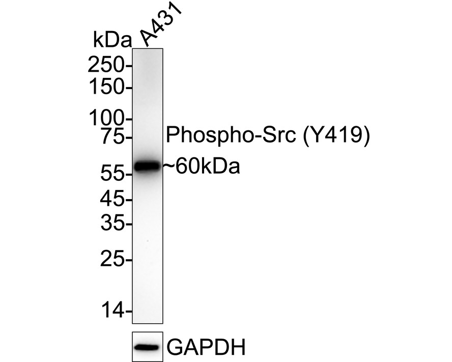
Western blot analysis of Phospho-Src (Y419) on A431 cell lysates with Rabbit anti-Phospho-Src (Y419) antibody at 1/1,000 dilution. Lysates/proteins at 20 µg/Lane. Exposure time: 59 seconds; 4-20% SDS-PAGE gel. Proteins were transferred to a PVDF membrane and blocked with 5% NFDM/TBST for 1 hour at room temperature. The primary antibody at 1/1,000 dilution was used in 5% NFDM/TBST at 4℃ overnight. Goat Anti-Rabbit IgG - HRP Secondary Antibody at 1/50,000 dilution was used for 1 hour at room temperature.ICC/IF
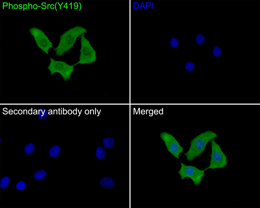
Immunocytochemistry analysis of A549 cells labeling Phospho-Src (Y419) with Rabbit anti-Phospho-Src (Y419) antibody at 1/50 dilution. Cells were fixed in 4% paraformaldehyde for 10 minutes at 37 ℃, permeabilized with 0.05% Triton X-100 in PBS for 20 minutes, and then blocked with 2% negative goat serum for 30 minutes at room temperature. Cells were then incubated with Rabbit anti-Phospho-Src (Y419) antibody at 1/50 dilution in 2% negative goat serum overnight at 4 ℃. Goat Anti-Rabbit IgG H&L (iFluor™ 488) was used as the secondary antibody at 1/1,000 dilution. PBS instead of the primary antibody was used as the secondary antibody only control. Nuclear DNA was labelled in blue with DAPI.FC
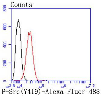
Flow cytometric analysis of Hela cells with Phospho-Src(Y419) antibody at 1/50 dilution (red) compared with an unlabelled control (cells without incubation with primary antibody; black). Alexa Fluor 488-conjugated goat anti rabbit IgG was used as the secondary antibody.| Product Name | Phospho-Src (Y419) Recombinant Rabbit Monoclonal Antibody |
|---|---|
| Antibody Type | Primary Antibodies |
| Product description | The Src family of protein tyrosine kinases, which includes Src, Lyn, Fyn, Yes, Lck, Blk, and Hck, are important in the regulation of growth and differentiation of eukaryotic cells. Src activity is regulated by tyrosine phosphorylation at two sites, but with opposing effects. While phosphorylation at Tyr416 in the activation loop of the kinase domain upregulates enzyme activity, phosphorylation at Tyr527 in the carboxy-terminal tail by Csk renders the enzyme less active. |
| Immunogen | Synthetic phospho-peptide corresponding to residues surrounding Tyr419 of Human Src aa 401-450 / 536. |
| Modification | p-Y419 |
| Clonality | monoclonal |
|---|---|
| Isotype | IgG |
| Host Species | Recombinant rabbit |
| Tested Applications | FCICC/IFWB |
| WB:1:1000 ICC/IF:1:50 FC:1:50-1:100 |
|
| Species Reactivity | Human |
| Concentration | 1mg/ml |
| Purification | Protein A |
| Alternative Names | ASV antibody Avian sarcoma virus antibody c SRC antibody CDNA FLJ14219 fis clone NT2RP3003800 highly similar to Rattus norvegicus tyrosine protein kinase pp60 c src mRNA antibody cSrc antibody EC 2.7.10.2 antibody Neuronal CSRC tyrosine specific protein kinase antibody Neuronal SRC antibody Oncogene SRC antibody OTTHUMP00000174476 antibody OTTHUMP00000174477 antibody p60 Src antibody p60-Src antibody p60Src antibody pp60c src antibody pp60c-src antibody pp60csrc antibody Proto oncogene tyrosine protein kinase Src antibody Proto-oncogene c-Src antibody Proto-oncogene tyrosine-protein kinase Src antibody Protooncogene SRC antibody Protooncogene SRC Rous sarcoma antibody Src antibody SRC Oncogene antibody SRC proto oncogene non receptor tyrosine kinase antibody SRC_HUMAN antibody SRC1 antibody Tyrosine kinase pp60c src antibody Tyrosine protein kinase SRC 1 antibody Tyrosine protein kinase SRC1 antibody v src avian sarcoma (Schmidt Ruppin A2) viral oncogene homolog antibody V src sarcoma (Schmidt Ruppin A 2) viral oncogene homolog (avian) antibody v src sarcoma (Schmidt Ruppin A 2) viral oncogene homolog avian antibody |
|---|---|
| Molecular Weight(MW) | 60 kDa |
| Cellular Localization | Cell membrane, Mitochondrion inner membrane, Nucleus, Cytoplasm. |
WB
Western blot analysis of Phospho-Src (Y419) on A431 cell lysates with Rabbit anti-Phospho-Src (Y419) antibody at 1/1,000 dilution. Lysates/proteins at 20 µg/Lane. Exposure time: 59 seconds; 4-20% SDS-PAGE gel. Proteins were transferred to a PVDF membrane and blocked with 5% NFDM/TBST for 1 hour at room temperature. The primary antibody at 1/1,000 dilution was used in 5% NFDM/TBST at 4℃ overnight. Goat Anti-Rabbit IgG - HRP Secondary Antibody at 1/50,000 dilution was used for 1 hour at room temperature.
ICC/IF
Immunocytochemistry analysis of A549 cells labeling Phospho-Src (Y419) with Rabbit anti-Phospho-Src (Y419) antibody at 1/50 dilution. Cells were fixed in 4% paraformaldehyde for 10 minutes at 37 ℃, permeabilized with 0.05% Triton X-100 in PBS for 20 minutes, and then blocked with 2% negative goat serum for 30 minutes at room temperature. Cells were then incubated with Rabbit anti-Phospho-Src (Y419) antibody at 1/50 dilution in 2% negative goat serum overnight at 4 ℃. Goat Anti-Rabbit IgG H&L (iFluor™ 488) was used as the secondary antibody at 1/1,000 dilution. PBS instead of the primary antibody was used as the secondary antibody only control. Nuclear DNA was labelled in blue with DAPI.
FC
Flow cytometric analysis of Hela cells with Phospho-Src(Y419) antibody at 1/50 dilution (red) compared with an unlabelled control (cells without incubation with primary antibody; black). Alexa Fluor 488-conjugated goat anti rabbit IgG was used as the secondary antibody.| Positive Control | Hela. |
|---|---|
| Application Notes | WB:1:1000 ICC/IF:1:50 FC:1:50-1:100 |
| Form | Liquid |
|---|---|
| Storage Instructions | Store at +4℃ after thawing. Aliquot store at -20℃ or -80℃. Avoid repeated freeze / thaw cycles. |
| Storage Buffer | 1*TBS (pH7.4), 1%BSA, 40%Glycerol. Preservative: 0.05% Sodium Azide. |


