WB
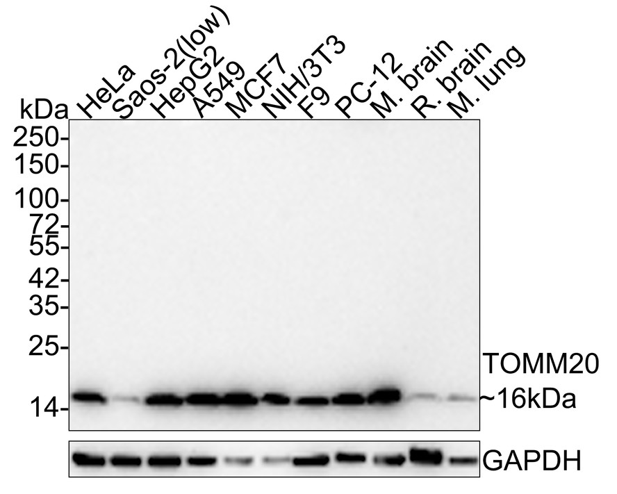
Western blot analysis of TOMM20 on different lysates with Rabbit anti-TOMM20 antibody at 1/5,000 dilution. Lane 1: HeLa cell lysate (10 µg/Lane), Lane 2: Saos-2 cell lysate (low expression) (10 µg/Lane), Lane 3: HepG2 cell lysate (10 µg/Lane), Lane 4: A549 cell lysate (10 µg/Lane), Lane 5: MCF7 cell lysate (10 µg/Lane), Lane 6: NIH/3T3 cell lysate (10 µg/Lane), Lane 7: F9 cell lysate (10 µg/Lane), Lane 8: PC-12 cell lysate (10 µg/Lane), Lane 9: Mouse brain tissue lysate (20 µg/Lane), Lane 10: Rat brain tissue lysate (20 µg/Lane), Lane 11: Rat lung tissue lysate (20 µg/Lane), Exposure time: 1 minute 22 seconds; 4-20% SDS-PAGE gel. Proteins were transferred to a PVDF membrane and blocked with 5% NFDM/TBST for 1 hour at room temperature. The primary antibody at 1/5,000 dilution was used in 5% NFDM/TBST at room temperature for 2 hours. Goat Anti-Rabbit IgG - HRP Secondary Antibody at 1/100,000 dilution was used for 1 hour at room temperature.IHC
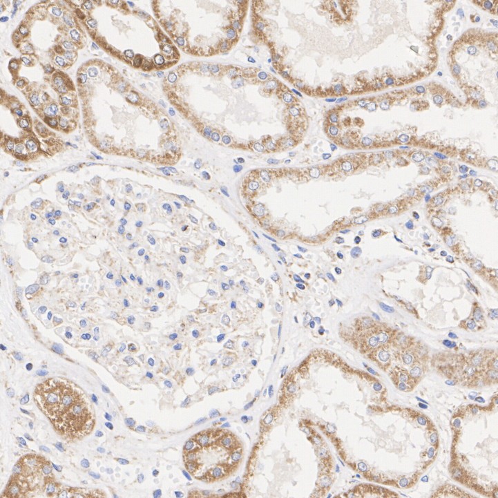
Immunohistochemical analysis of paraffin-embedded human kidney tissue with Rabbit anti-TOMM20 antibody at 1/500 dilution. The section was pre-treated using heat mediated antigen retrieval with Tris-EDTA buffer (pH 9.0) for 20 minutes. The tissues were blocked in 1% BSA for 20 minutes at room temperature, washed with ddH2O and PBS, and then probed with the primary antibody at 1/500 dilution for 1 hour at room temperature. The detection was performed using an HRP conjugated compact polymer system. DAB was used as the chromogen. Tissues were counterstained with hematoxylin and mounted with DPX.IHC
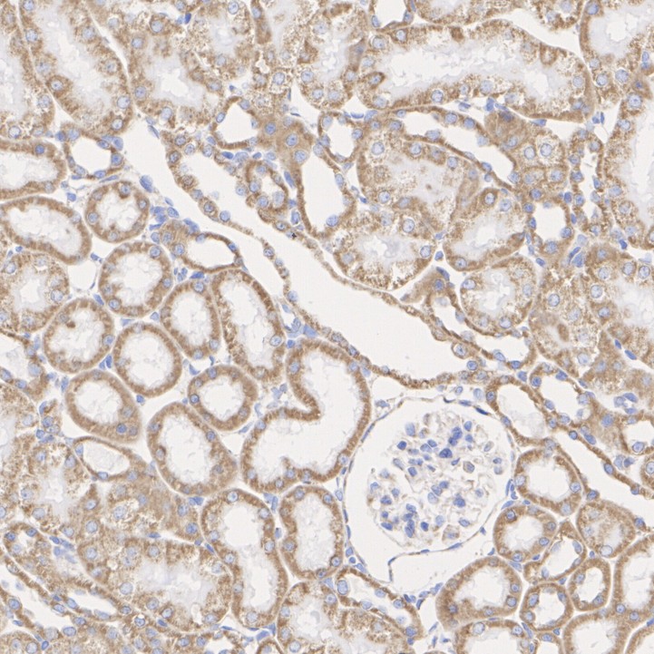
Immunohistochemical analysis of paraffin-embedded rat kidney tissue with Rabbit anti-TOMM20 antibody at 1/500 dilution. The section was pre-treated using heat mediated antigen retrieval with Tris-EDTA buffer (pH 9.0) for 20 minutes. The tissues were blocked in 1% BSA for 20 minutes at room temperature, washed with ddH2O and PBS, and then probed with the primary antibody at 1/500 dilution for 1 hour at room temperature. The detection was performed using an HRP conjugated compact polymer system. DAB was used as the chromogen. Tissues were counterstained with hematoxylin and mounted with DPX.IHC
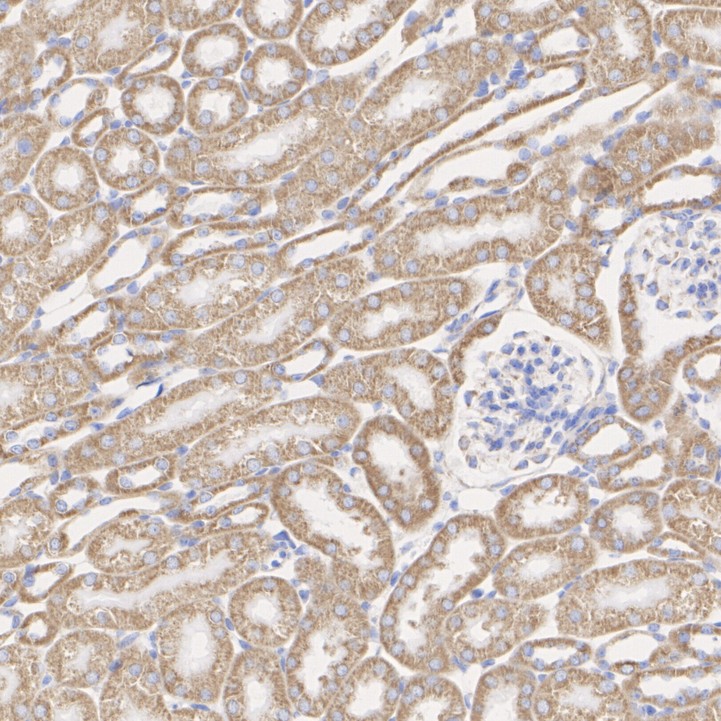
Immunohistochemical analysis of paraffin-embedded mouse kidney tissue with Rabbit anti-TOMM20 antibody at 1/500 dilution. The section was pre-treated using heat mediated antigen retrieval with Tris-EDTA buffer (pH 9.0) for 20 minutes. The tissues were blocked in 1% BSA for 20 minutes at room temperature, washed with ddH2O and PBS, and then probed with the primary antibody at 1/500 dilution for 1 hour at room temperature. The detection was performed using an HRP conjugated compact polymer system. DAB was used as the chromogen. Tissues were counterstained with hematoxylin and mounted with DPX.ICC/IF
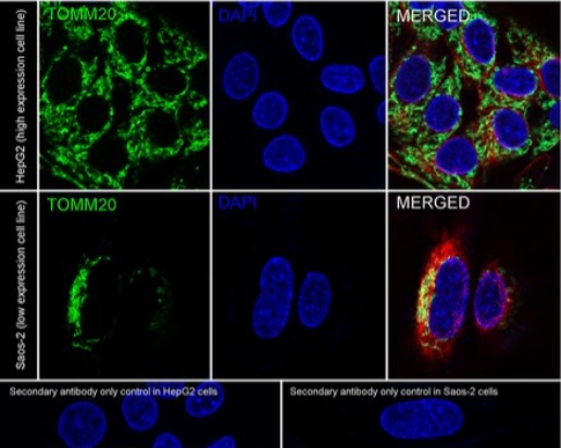
Immunocytochemistry analysis of HepG2 (high expression) and Saos-2 (low expression) labeling TOMM20 with Rabbit anti-TOMM20 antibody at 1/1,000 dilution. Cells were fixed in 4% paraformaldehyde for 20 minutes at room temperature, permeabilized with 0.1% Triton X-100 in PBS for 5 minutes at room temperature, then blocked with 1% BSA in 10% negative goat serum for 1 hour at room temperature. Cells were then incubated with Rabbit anti-TOMM20 antibody at 1/1,000 dilution in 1% BSA in PBST overnight at 4 ℃. Goat Anti-Rabbit IgG H&L (488) was used as the secondary antibody at 1/1,000 dilution. PBS instead of the primary antibody was used as the secondary antibody only control. Nuclear DNA was labelled in blue with DAPI. Beta tubulin (red) was stained at 1/100 dilution overnight at +4℃. Goat Anti-Mouse IgG H&L (594) was used as the secondary antibody at 1/1,000 dilution.FC
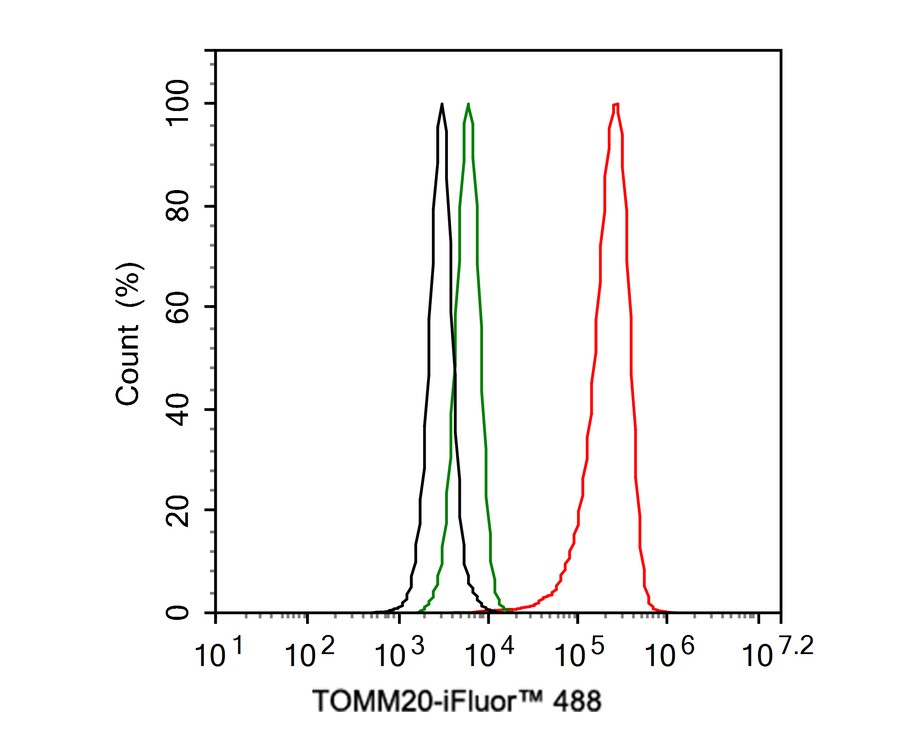
Flow cytometric analysis of HeLa cells labeling TOMM20. Cells were fixed and permeabilized. Then stained with the primary antibody (1μg/mL) (red) compared with Rabbit IgG Isotype Control (green). After incubation of the primary antibody at +4℃ for an hour, the cells were stained with a iFluor™ 488 conjugate-Goat anti-Rabbit IgG Secondary antibody at 1/1,000 dilution for 30 minutes at +4℃. Unlabelled sample was used as a control (cells without incubation with primary antibody; black).IP
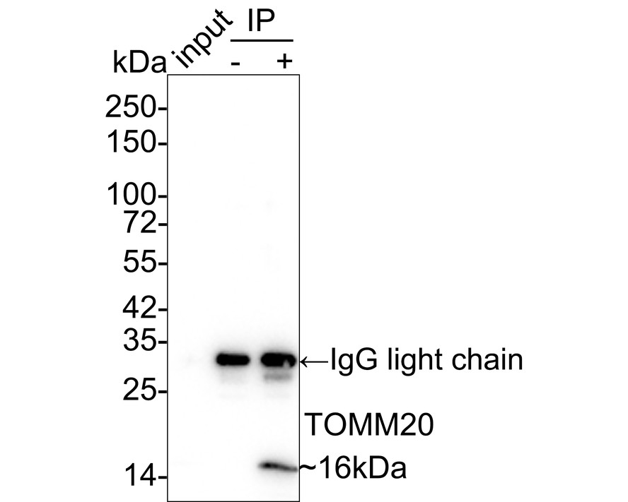
TOMM20 was immunoprecipitated in 0.2mg HeLa cell lysate with Rabbit anti-TOMM20 antibody at 2 µg/25 µl agarose. Western blot was performed from the immunoprecipitate using Rabbit anti-TOMM20 antibody at 1/1,000 dilution. Anti-Rabbit IgG for IP Nano-secondary antibody at 1/5,000 dilution was used for 1 hour at room temperature. Lane 1: HeLa cell lysate (input), Lane 2: Rabbit IgG instead of Rabbit anti-TOMM20 antibody in HeLa cell lysate, Lane 3: Rabbit anti-TOMM20 antibody IP in HeLa cell lysate, Blocking/Dilution buffer: 5% NFDM/TBST, Exposure time: 1 minute 2 seconds.| Product Name | TOMM20 Recombinant Rabbit Monoclonal Antibody |
|---|---|
| Antibody Type | Primary Antibodies |
| Product description | The mitochondrial preprotein translocases of the outer membrane (Tom) is a multisubunit protein complex that facilitates the import of nucleus-encoded precursor proteins across the mitochondrial outer membrane. The Tom machinery consists of import receptors for the initial binding of cytosolically synthesized preproteins and a general import pore (GIP) for the membrane translocation of various preproteins into the mitochondria. The import receptors include Tom20 and Tom22, which form a heteromeric receptor complex that initiates the insertion of newly synthesized proteins into the outer membrane and then directs the precursor protein into the GIP. In yeast, Tom22 is the essential component of the import receptor complex as it functions as both a receptor for the preproteins and serves as a docking point for both Tom20 and the GIP. Tom22 directly associates with Tom40, the major component of the GIP, and thereby forms a stable interaction between the two core complexes to facilitate the fluid movement of preproteins into the mitochondria. The insertion of Tom40 into the Tom machinery requires the initial binding of Tom40 to Tom20 and leads to the efficient incorporation of Tom40 precursors into preexisting Tom complexes. |
| Immunogen | Recombinant protein within Human TOMM20 aa 1-145 / 145. |
| Clonality | Monoclonal |
|---|---|
| Isotype | IgG |
| Host Species | Recombinant rabbit |
| Tested Applications | FCICC/IFIHCIPWB |
| WB:1:5000-1:20000 IHC1:500-1:1000ICC/IF:1:500-1:1000 FC:1:1000 IP:Use at an assay dependent concentration. |
|
| Species Reactivity | HumanMouseRat |
| Concentration | 1mg/ml |
| Purification | Protein A |
| Alternative Names | KIAA0016 antibody MAS20 antibody MGC117367 antibody Mitochondrial 20 kDa outer membrane protein antibody Mitochondrial import receptor subunit TOM20 homolog antibody MOM19 antibody Outer mitochondrial membrane receptor Tom20 antibody TOM20 antibody TOM20_HUMAN antibody TOMM20 antibody Translocase of outer mitochondrial membrane 20 homolog (yeast) antibody Translocase of outer mitochondrial membrane 20 homolog type II antibody |
|---|---|
| Molecular Weight(MW) | 16 kDa |
| Cellular Localization | Mitochondrion outer membrane. |
WB
Western blot analysis of TOMM20 on different lysates with Rabbit anti-TOMM20 antibody at 1/5,000 dilution. Lane 1: HeLa cell lysate (10 µg/Lane), Lane 2: Saos-2 cell lysate (low expression) (10 µg/Lane), Lane 3: HepG2 cell lysate (10 µg/Lane), Lane 4: A549 cell lysate (10 µg/Lane), Lane 5: MCF7 cell lysate (10 µg/Lane), Lane 6: NIH/3T3 cell lysate (10 µg/Lane), Lane 7: F9 cell lysate (10 µg/Lane), Lane 8: PC-12 cell lysate (10 µg/Lane), Lane 9: Mouse brain tissue lysate (20 µg/Lane), Lane 10: Rat brain tissue lysate (20 µg/Lane), Lane 11: Rat lung tissue lysate (20 µg/Lane), Exposure time: 1 minute 22 seconds; 4-20% SDS-PAGE gel. Proteins were transferred to a PVDF membrane and blocked with 5% NFDM/TBST for 1 hour at room temperature. The primary antibody at 1/5,000 dilution was used in 5% NFDM/TBST at room temperature for 2 hours. Goat Anti-Rabbit IgG - HRP Secondary Antibody at 1/100,000 dilution was used for 1 hour at room temperature.
IHC
Immunohistochemical analysis of paraffin-embedded human kidney tissue with Rabbit anti-TOMM20 antibody at 1/500 dilution. The section was pre-treated using heat mediated antigen retrieval with Tris-EDTA buffer (pH 9.0) for 20 minutes. The tissues were blocked in 1% BSA for 20 minutes at room temperature, washed with ddH2O and PBS, and then probed with the primary antibody at 1/500 dilution for 1 hour at room temperature. The detection was performed using an HRP conjugated compact polymer system. DAB was used as the chromogen. Tissues were counterstained with hematoxylin and mounted with DPX.
IHC
Immunohistochemical analysis of paraffin-embedded rat kidney tissue with Rabbit anti-TOMM20 antibody at 1/500 dilution. The section was pre-treated using heat mediated antigen retrieval with Tris-EDTA buffer (pH 9.0) for 20 minutes. The tissues were blocked in 1% BSA for 20 minutes at room temperature, washed with ddH2O and PBS, and then probed with the primary antibody at 1/500 dilution for 1 hour at room temperature. The detection was performed using an HRP conjugated compact polymer system. DAB was used as the chromogen. Tissues were counterstained with hematoxylin and mounted with DPX.
IHC
Immunohistochemical analysis of paraffin-embedded mouse kidney tissue with Rabbit anti-TOMM20 antibody at 1/500 dilution. The section was pre-treated using heat mediated antigen retrieval with Tris-EDTA buffer (pH 9.0) for 20 minutes. The tissues were blocked in 1% BSA for 20 minutes at room temperature, washed with ddH2O and PBS, and then probed with the primary antibody at 1/500 dilution for 1 hour at room temperature. The detection was performed using an HRP conjugated compact polymer system. DAB was used as the chromogen. Tissues were counterstained with hematoxylin and mounted with DPX.
ICC/IF
Immunocytochemistry analysis of HepG2 (high expression) and Saos-2 (low expression) labeling TOMM20 with Rabbit anti-TOMM20 antibody at 1/1,000 dilution. Cells were fixed in 4% paraformaldehyde for 20 minutes at room temperature, permeabilized with 0.1% Triton X-100 in PBS for 5 minutes at room temperature, then blocked with 1% BSA in 10% negative goat serum for 1 hour at room temperature. Cells were then incubated with Rabbit anti-TOMM20 antibody at 1/1,000 dilution in 1% BSA in PBST overnight at 4 ℃. Goat Anti-Rabbit IgG H&L (488) was used as the secondary antibody at 1/1,000 dilution. PBS instead of the primary antibody was used as the secondary antibody only control. Nuclear DNA was labelled in blue with DAPI. Beta tubulin (red) was stained at 1/100 dilution overnight at +4℃. Goat Anti-Mouse IgG H&L (594) was used as the secondary antibody at 1/1,000 dilution.
FC
Flow cytometric analysis of HeLa cells labeling TOMM20. Cells were fixed and permeabilized. Then stained with the primary antibody (1μg/mL) (red) compared with Rabbit IgG Isotype Control (green). After incubation of the primary antibody at +4℃ for an hour, the cells were stained with a iFluor™ 488 conjugate-Goat anti-Rabbit IgG Secondary antibody at 1/1,000 dilution for 30 minutes at +4℃. Unlabelled sample was used as a control (cells without incubation with primary antibody; black).
IP
TOMM20 was immunoprecipitated in 0.2mg HeLa cell lysate with Rabbit anti-TOMM20 antibody at 2 µg/25 µl agarose. Western blot was performed from the immunoprecipitate using Rabbit anti-TOMM20 antibody at 1/1,000 dilution. Anti-Rabbit IgG for IP Nano-secondary antibody at 1/5,000 dilution was used for 1 hour at room temperature. Lane 1: HeLa cell lysate (input), Lane 2: Rabbit IgG instead of Rabbit anti-TOMM20 antibody in HeLa cell lysate, Lane 3: Rabbit anti-TOMM20 antibody IP in HeLa cell lysate, Blocking/Dilution buffer: 5% NFDM/TBST, Exposure time: 1 minute 2 seconds.| Application Notes | WB:1:5000-1:20000 IHC1:500-1:1000ICC/IF:1:500-1:1000 FC:1:1000 IP:Use at an assay dependent concentration. |
|---|
| Form | Liquid |
|---|---|
| Storage Instructions | Store at +4℃ after thawing. Aliquot store at -20℃ or -80℃. Avoid repeated freeze / thaw cycles. |
| Storage Buffer | 1*TBS (pH7.4), 1%BSA, 40%Glycerol. Preservative: 0.05% Sodium Azide. |


