Application
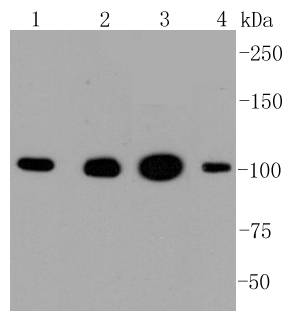
Fig1: Western blot analysis of Hexokinase 1 on different lysates using anti-Hexokinase 1 antibody at 1/1,000 dilution. Positive control: Lane 1: Hela Lane 2: 293 Lane 3: MCF-7 Lane 4: HepG2Application
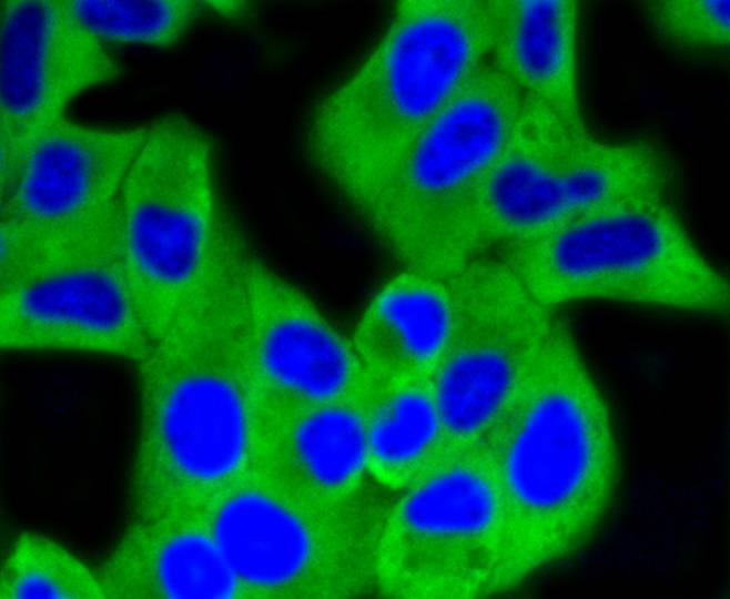
Fig2: ICC staining Hexokinase 1 in Hela cells (green). The nuclear counter stain is DAPI (blue). Cells were fixed in paraformaldehyde, permeabilised with 0.25% Triton X100/PBS.Application
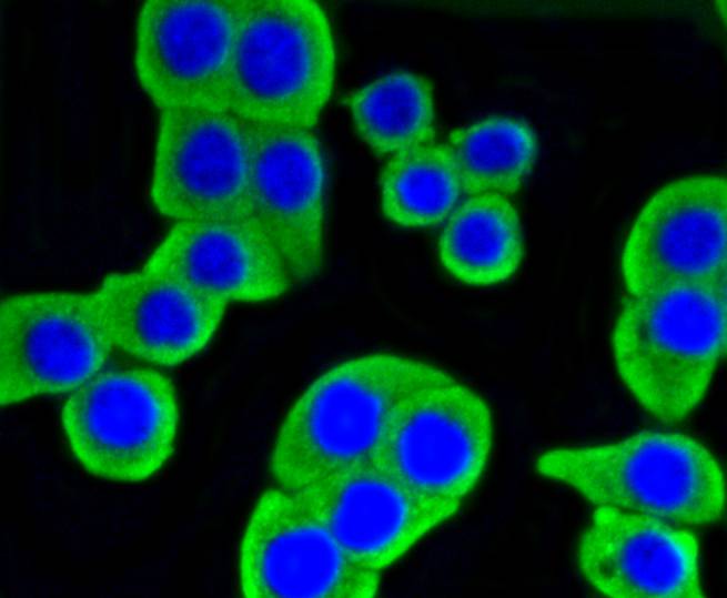
Fig3: ICC staining Hexokinase 1 in CRC cells (green). The nuclear counter stain is DAPI (blue). Cells were fixed in paraformaldehyde, permeabilised with 0.25% Triton X100/PBS.Application
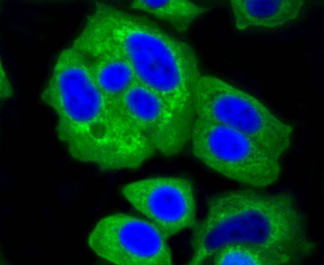
Fig4: ICC staining Hexokinase 1 in MCF-7 cells (green). The nuclear counter stain is DAPI (blue). Cells were fixed in paraformaldehyde, permeabilised with 0.25% Triton X100/PBS.Application
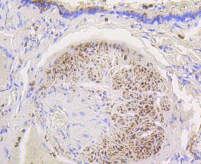
Fig5: Immunohistochemical analysis of paraffin-embedded human lung tissue using anti-Hexokinase 1 antibody. Counter stained with hematoxylin.Application
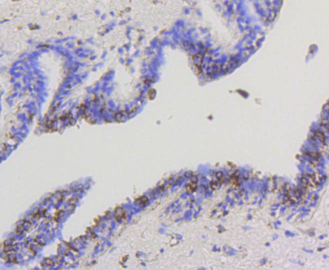
Fig6: Immunohistochemical analysis of paraffin-embedded human breast carcinoma tissue using anti-Hexokinase 1 antibody. Counter stained with hematoxylin.Application
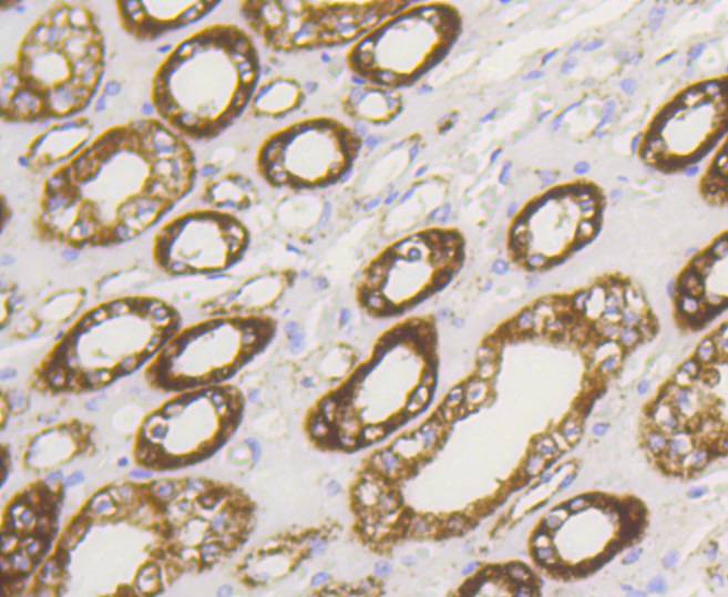
Fig7: Immunohistochemical analysis of paraffin-embedded human kidney tissue using anti-Hexokinase 1 antibody. Counter stained with hematoxylin.Application
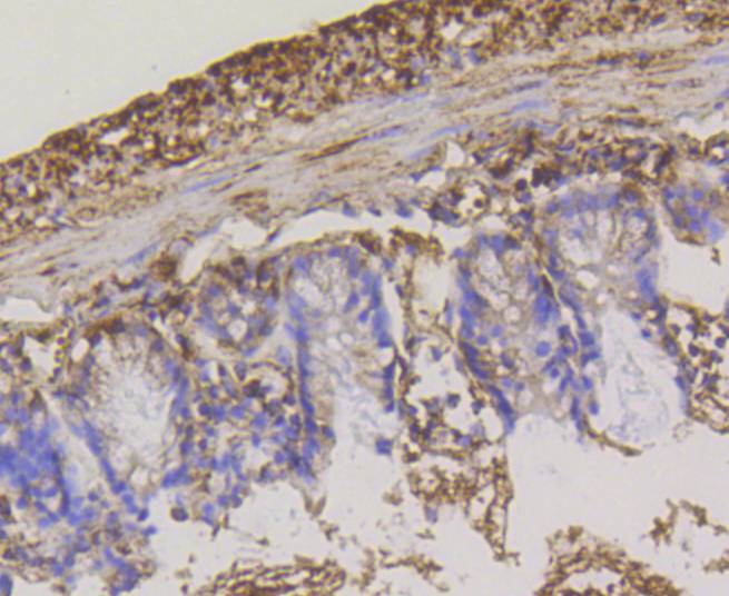
Fig8: Immunohistochemical analysis of paraffin-embedded mouse colon tissue using anti-Hexokinase 1 antibody. Counter stained with hematoxylin.Application
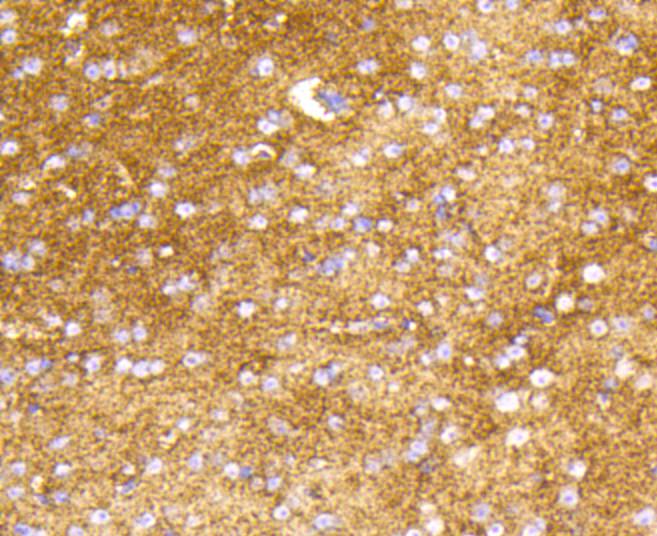
Fig9: Immunohistochemical analysis of paraffin-embedded mouse brain tissue using anti-Hexokinase 1 antibody. Counter stained with hematoxylin.Application
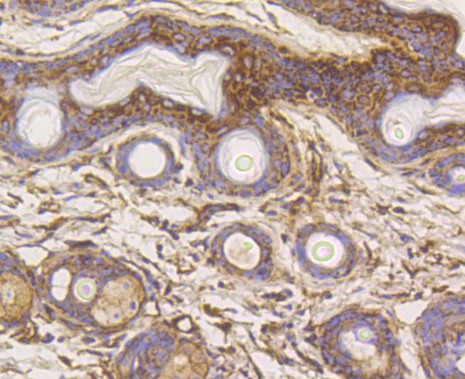
Fig10: Immunohistochemical analysis of paraffin-embedded mouse skin tissue using anti-Hexokinase 1 antibody. Counter stained with hematoxylin.Application
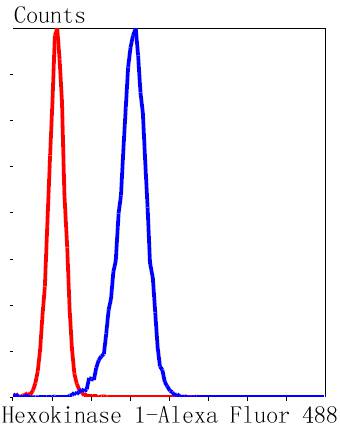
Fig11: Flow cytometric analysis of K562 cells with Hexokinase 1 antibody at 1/50 dilution (blue) compared with an unlabelled control (cells without incubation with primary antibody; red). Alexa Fluor 488-conjugated goat anti rabbit IgG was used as the secondary antibody| Product Name | Hexokinase 1 [ST47-05] |
|---|---|
| Antibody Type | Primary Antibodies |
| Antigen Alias | BB404130 antibody Brain form hexokinase antibody dea antibody DrHXK1 antibody EC 2.7.1.1 antibody Glycolytic enzyme antibody HEXOKIN antibody hexokinase I antibody Hexokinase PI antibody Hexokinase type I antibody Hexokinase, tumor isozyme antibody Hexokinase-1 antibody Hexokinase-A antibody HK I antibody HK1 antibody HK1 tb antibody Hk1-s antibody HK1-ta antibody HK1-tb antibody HK1-tc antibody HKD antibody HKI antibody HMSNR antibody HXK1 antibody HXK1_HUMAN antibody im:7148527 antibody mHk1-s antibody wu:fc09d08 antibody wu:fc16e02 antibody wu:fc21e02 antibody wu:fq14b11 antibody zgc:55790 antibody zgc:77618 antibody |
| Product description | The hexokinases utilize Mg-ATP as a phosphoryl donor to catalyze the first step of intracellular glucose metabolism, the conversion of glucose to glucose-6-phosphate. Four hexokinase isoenzymes have been identified, including hexokinase I (HXK I), hexokinase II (HXK II), hexokinase III (HXK III) and hexokinase IV (HXK IV, also designated glucokinase or GCK). Hexokinases I-III each contain an N-terminal cluster of hydrophobic amino acids. Glucokinase lacks the N-terminal hydrophobic cluster. The hydrophobic cluster is thought to be necessary for membrane binding. This is substantiated by the finding that glucokinase has lower affinity for glucose than do the other hexokinases. HXK I has been shown to be expressed in brain, kidney and heart tissues as well as in hepatoma cell lines. HXK II is involved in the uptake and utilization of glucose by adipose and skeletal tissues. Of the hexokinases, HXK III has the highest affinity for glucose. Glucokinase is expressed in pancreatic beta cells where it functions as a glucose sensor, determining the “set point” for insulin secretion. |
| Immunogen | recombinant protein |
| Clonality | Monoclonal |
|---|---|
| Isotype | IgG |
| Host Species | Recombinant rabbit |
| Tested Applications | WBICC/IFIHCIPFC |
| WB:1:1,000-1:2,000 ICC:1:50-1:200 IHC:1:50-1:200 FC:1:50-1:100 |
|
| Species Reactivity | HumanMouseRat |
| Concentration | 1mg/ml |
| Alternative Names | BB404130 antibody
Brain form hexokinase antibody
dea antibody
DrHXK1 antibody
EC 2.7.1.1 antibody
Glycolytic enzyme antibody
HEXOKIN antibody
hexokinase I antibody
Hexokinase PI antibody
Hexokinase type I antibody
Hexokinase tumor isozyme antibody Hexokinase-1 antibody Hexokinase-A antibody HK I antibody HK1 antibody HK1 tb antibody Hk1-s antibody HK1-ta antibody HK1-tb antibody HK1-tc antibody HKD antibody HKI antibody HMSNR antibody HXK1 antibody HXK1_HUMAN antibody im:7148527 antibody mHk1-s antibody wu:fc09d08 antibody wu:fc16e02 antibody wu:fc21e02 antibody wu:fq14b11 antibody zgc:55790 antibody zgc:77618 antibody |
|---|---|
| Molecular Weight(MW) | 102 kDa |
| Cellular Localization | Mitochondrion outer membrane. |
Application
Fig1: Western blot analysis of Hexokinase 1 on different lysates using anti-Hexokinase 1 antibody at 1/1,000 dilution. Positive control: Lane 1: Hela Lane 2: 293 Lane 3: MCF-7 Lane 4: HepG2
Application
Fig2: ICC staining Hexokinase 1 in Hela cells (green). The nuclear counter stain is DAPI (blue). Cells were fixed in paraformaldehyde, permeabilised with 0.25% Triton X100/PBS.
Application
Fig3: ICC staining Hexokinase 1 in CRC cells (green). The nuclear counter stain is DAPI (blue). Cells were fixed in paraformaldehyde, permeabilised with 0.25% Triton X100/PBS.
Application
Fig4: ICC staining Hexokinase 1 in MCF-7 cells (green). The nuclear counter stain is DAPI (blue). Cells were fixed in paraformaldehyde, permeabilised with 0.25% Triton X100/PBS.
Application
Fig5: Immunohistochemical analysis of paraffin-embedded human lung tissue using anti-Hexokinase 1 antibody. Counter stained with hematoxylin.
Application
Fig6: Immunohistochemical analysis of paraffin-embedded human breast carcinoma tissue using anti-Hexokinase 1 antibody. Counter stained with hematoxylin.
Application
Fig7: Immunohistochemical analysis of paraffin-embedded human kidney tissue using anti-Hexokinase 1 antibody. Counter stained with hematoxylin.
Application
Fig8: Immunohistochemical analysis of paraffin-embedded mouse colon tissue using anti-Hexokinase 1 antibody. Counter stained with hematoxylin.
Application
Fig9: Immunohistochemical analysis of paraffin-embedded mouse brain tissue using anti-Hexokinase 1 antibody. Counter stained with hematoxylin.
Application
Fig10: Immunohistochemical analysis of paraffin-embedded mouse skin tissue using anti-Hexokinase 1 antibody. Counter stained with hematoxylin.
Application
Fig11: Flow cytometric analysis of K562 cells with Hexokinase 1 antibody at 1/50 dilution (blue) compared with an unlabelled control (cells without incubation with primary antibody; red). Alexa Fluor 488-conjugated goat anti rabbit IgG was used as the secondary antibody| Positive Control | CRC, MCF-7, Hela, HepG2, mouse colon tissue, human lung tissue, mouse brain tissue, human breast carcinoma tissue, mouse skin tissue, human kidney tissue. |
|---|---|
| Application Notes | WB:1:1,000-1:2,000 ICC:1:50-1:200 IHC:1:50-1:200 FC:1:50-1:100 |
| Form | Liquid |
|---|---|
| Storage Instructions | Store at +4℃ after thawing. Aliquot store at -20℃ or -80℃. Avoid repeated freeze / thaw cycles. |
| Storage Buffer | 1*TBS (pH7.4), 1%BSA, 40%Glycerol. Preservative: 0.05% Sodium Azide. |


