Application
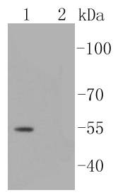
Fig1: Western blot analysis of Phospho-Smad3(S423/S425) on different lysates using anti-Phospho-Smad3(S423/S425) antibody at 1/1,000 dilution.Positive control:Lane 1: A431 cell lysates-treated with TGF βLane 2: A431 cell lysates-untreatedApplication
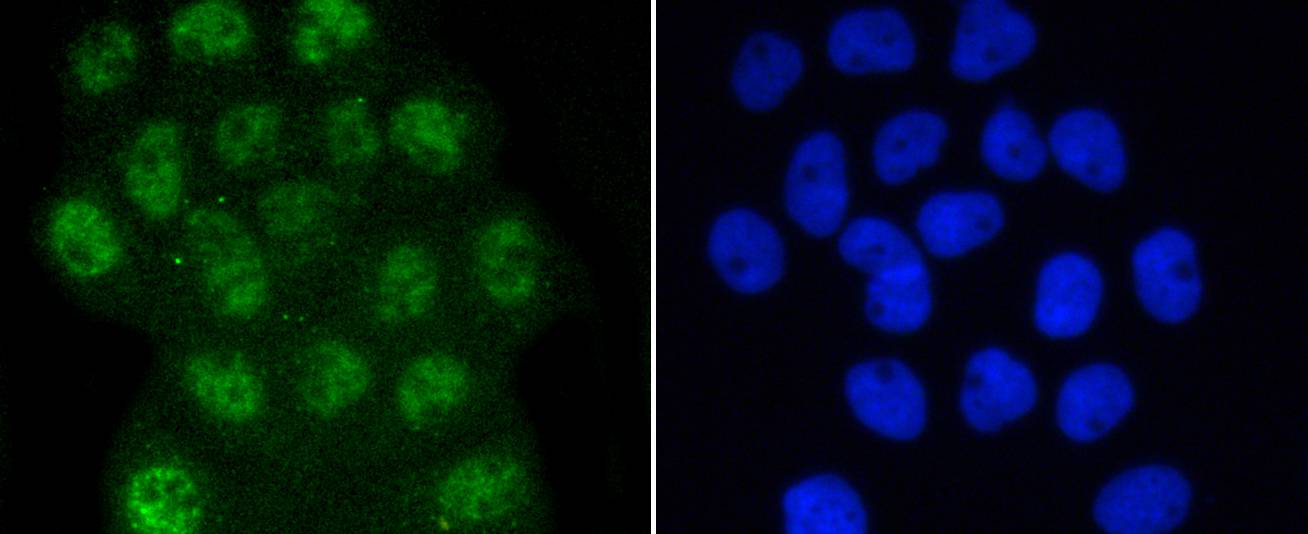
Fig2: ICC staining Phospho-Smad3(S423/S425) in A431 cells (green). The nuclear counter stain is DAPI (blue). Cells were fixed in paraformaldehyde, permeabilised with 0.25% Triton X100/PBS.Application
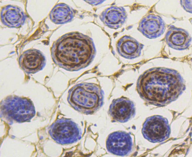
Fig3: Immunohistochemical analysis of paraffin-embedded mouse skin tissue using anti-Phospho-Smad3(S423/S425) antibody. Counter stained with hematoxylin.| Product Name | Phospho-Smad3(S423/S425) [ST0493] |
|---|---|
| Antibody Type | Primary Antibodies |
| Antigen Alias | DKFZP586N0721 antibody DKFZp686J10186 antibody hMAD 3 antibody hMAD-3 antibody hSMAD3 antibody HSPC193 antibody HST17436 antibody JV15 2 antibody JV15-2 antibody JV152 antibody LDS1C antibody LDS3 antibody MAD (mothers against decapentaplegic Drosophila) homolog 3 antibody MAD homolog 3 antibody Mad homolog JV15 2 antibody Mad protein homolog antibody MAD, mothers against decapentaplegic homolog 3 antibody Mad3 antibody MADH 3 antibody MADH3 antibody MGC60396 antibody Mothers against decapentaplegic homolog 3 antibody Mothers against DPP homolog 3 antibody SMA and MAD related protein 3 antibody SMAD 3 antibody SMAD antibody SMAD family member 3 antibody SMAD, mothers against DPP homolog 3 antibody Smad3 antibody SMAD3_HUMAN antibody |
| Product description | nst dpp (Mad) have been implicated as downstream effectors of TGFβ/BMP signaling. Smad1 (also designated Madr1 or JV4-1), Smad5 and mammalian Smad8 (also designated Smad9 or MADH6) are effectors of BMP2 and BMP4 function while Smad2 (also designated Madr2 or JV18-1) and Smad3 are involved in TGFβ and activin-mediated growth modulation. Smad4 (also designated DPC4) has been shown to mediate all of the above activities through interaction with various Smad family members. Smad6 and Smad7 regulate the response to activin/TGFβ signaling by interfering with TGFβ-mediated phosphorylation of other Smad family members. Human Smad3 is subject to phosphorylation by TGFβ on specific amino acid residues, including Ser 208. |
| Immunogen | Synthetic phospho-peptide corresponding to residues surrounding Ser423 and 425 of human Smad3. |
| Modification | p-S423/S425 |
| Clonality | Monoclonal |
|---|---|
| Isotype | IgG |
| Host Species | Recombinant rabbit |
| Tested Applications | WBICC/IFIHC |
| WB:1:1,000 ICC:1:50-1:200 IHC:1:50-1:200 |
|
| Species Reactivity | HumanMouse |
| Concentration | 1mg/ml |
| Alternative Names | DKFZP586N0721 antibody
DKFZp686J10186 antibody
hMAD 3 antibody
hMAD-3 antibody
hSMAD3 antibody
HSPC193 antibody
HST17436 antibody
JV15 2 antibody
JV15-2 antibody
JV152 antibody
LDS1C antibody
LDS3 antibody
MAD (mothers against decapentaplegic Drosophila) homolog 3 antibody
MAD homolog 3 antibody
Mad homolog JV15 2 antibody
Mad protein homolog antibody
MAD mothers against decapentaplegic homolog 3 antibody Mad3 antibody MADH 3 antibody MADH3 antibody MGC60396 antibody Mothers against decapentaplegic homolog 3 antibody Mothers against DPP homolog 3 antibody SMA and MAD related protein 3 antibody SMAD 3 antibody SMAD antibody SMAD family member 3 antibody SMAD mothers against DPP homolog 3 antibody Smad3 antibody SMAD3_HUMAN antibody |
|---|---|
| Molecular Weight(MW) | 55 kDa |
| Cellular Localization | Cytoplasm, Nucleus. |
Application
Fig1: Western blot analysis of Phospho-Smad3(S423/S425) on different lysates using anti-Phospho-Smad3(S423/S425) antibody at 1/1,000 dilution.Positive control:Lane 1: A431 cell lysates-treated with TGF βLane 2: A431 cell lysates-untreated
Application
Fig2: ICC staining Phospho-Smad3(S423/S425) in A431 cells (green). The nuclear counter stain is DAPI (blue). Cells were fixed in paraformaldehyde, permeabilised with 0.25% Triton X100/PBS.
Application
Fig3: Immunohistochemical analysis of paraffin-embedded mouse skin tissue using anti-Phospho-Smad3(S423/S425) antibody. Counter stained with hematoxylin.| Positive Control | A431 cell lysates-treated with TGF β, mouse skin tissue. |
|---|---|
| Application Notes | WB:1:1,000 ICC:1:50-1:200 IHC:1:50-1:200 |
| Form | Liquid |
|---|---|
| Storage Instructions | Store at +4℃ after thawing. Aliquot store at -20℃ or -80℃. Avoid repeated freeze / thaw cycles. |
| Storage Buffer | 1*TBS (pH7.4), 1%BSA, 40%Glycerol. Preservative: 0.05% Sodium Azide. |


