WB
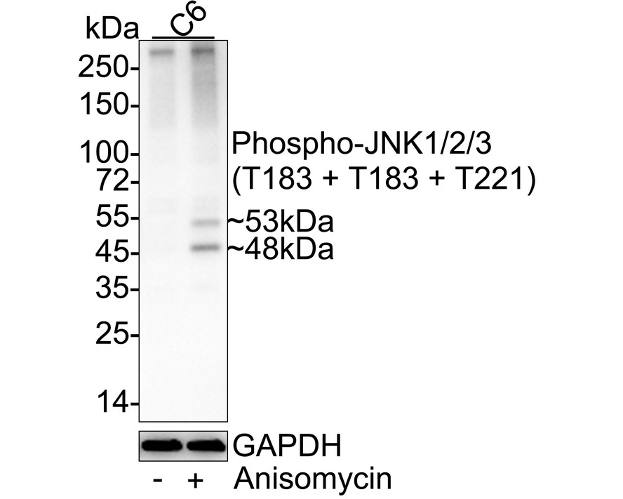
Western blot analysis of Phospho-JNK1/2/3 (T183 + T183 + T221) on different lysates with Rabbit anti-Phospho-JNK1/2/3 (T183 + T183 + T221) antibody at 1/2,000 dilution. Lane 1: C6 cell lysate, Lane 2: C6 treated with 25µg/mL Anisomycin for 30 minutes cell lysate. Lysates/proteins at 20 µg/Lane. Exposure time: 2 minutes 6 seconds; 4-20% SDS-PAGE gel. Proteins were transferred to a PVDF membrane and blocked with 5% NFDM/TBST for 1 hour at room temperature. The primary antibody at 1/2,000 dilution was used in 5% NFDM/TBST at 4℃ overnight. Goat Anti-Rabbit IgG - HRP Secondary Antibody at 1/50,000 dilution was used for 1 hour at room temperature.WB
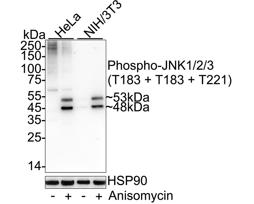
Western blot analysis of Phospho-JNK1/2/3 (T183 + T183 + T221) on different lysates with Rabbit anti-Phospho-JNK1/2/3 (T183 + T183 + T221) antibody at 1/2,000 dilution. Lane 1: HeLa cell lysate, Lane 2: HeLa treated with 25μg/mL Anisomycin for 30 minutes cell lysate, Lane 3: NIH/3T3 cell lysate, Lane 4: NIH/3T3 treated with 25μg/mL Anisomycin for 30 minutes cell lysate, Lysates/proteins at 20 µg/Lane. Exposure time: 59 seconds; 4-20% SDS-PAGE gel. Proteins were transferred to a PVDF membrane and blocked with 5% NFDM/TBST for 1 hour at room temperature. The primary antibody at 1/2,000 dilution was used in 5% NFDM/TBST at 4℃ overnight. Goat Anti-Rabbit IgG - HRP Secondary Antibody at 1/50,000 dilution was used for 1 hour at room temperature.IHC
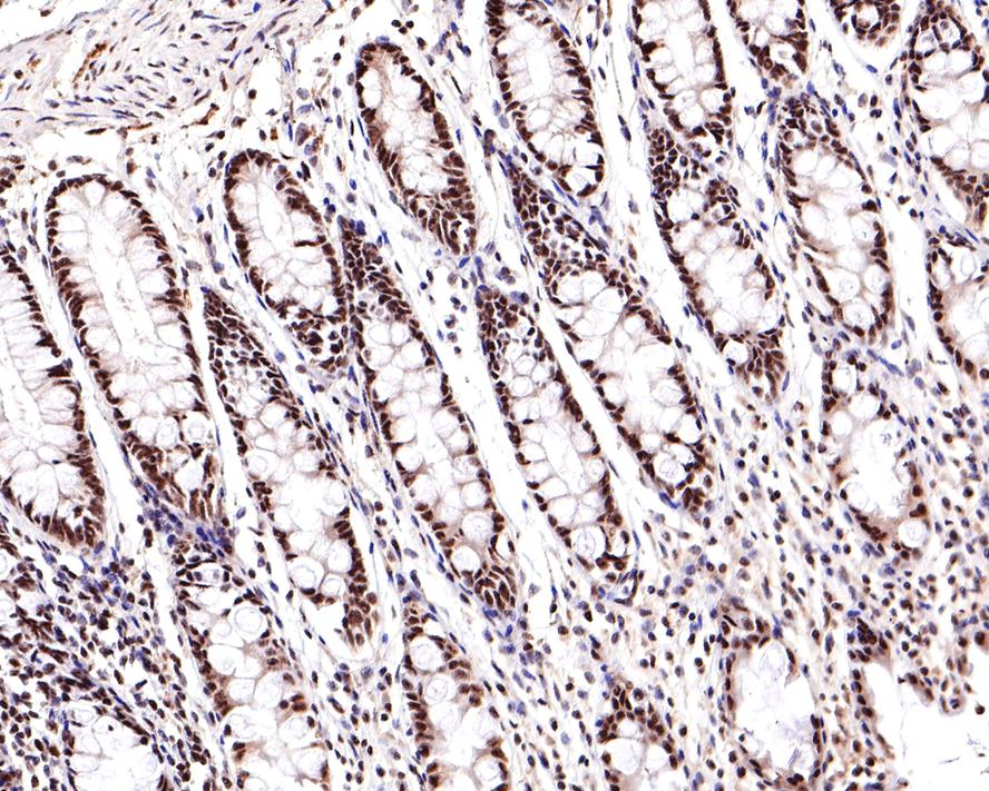
Immunohistochemical analysis of paraffin-embedded human colon tissue with Rabbit anti-Phospho-JNK1/2/3 (T183 + T183 + T221) antibody at 1/400 dilution. The section was pre-treated using heat mediated antigen retrieval with sodium citrate buffer (pH 6.0) for 2 minutes. The tissues were blocked in 1% BSA for 20 minutes at room temperature, washed with ddH2O and PBS, and then probed with the primary antibody at 1/400 dilution for 1 hour at room temperature. The detection was performed using an HRP conjugated compact polymer system. DAB was used as the chromogen. Tissues were counterstained with hematoxylin and mounted with DPX.IHC
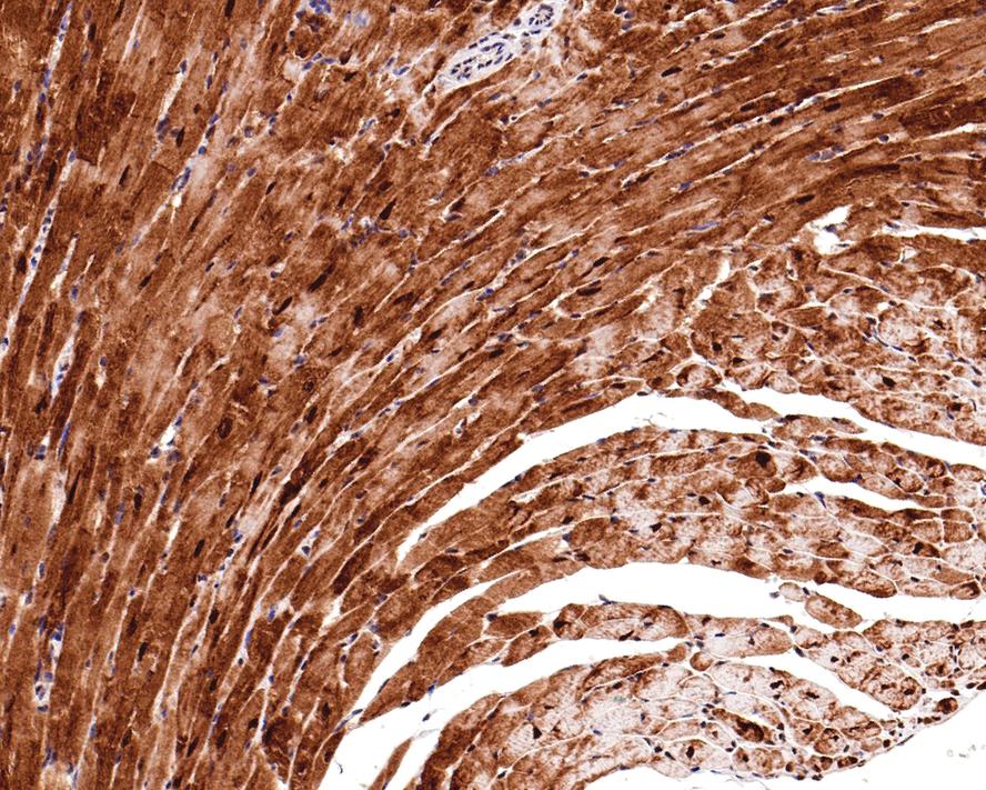
Immunohistochemical analysis of paraffin-embedded mouse heart tissue with Rabbit anti-Phospho-JNK1/2/3 (T183 + T183 + T221) antibody at 1/400 dilution. The section was pre-treated using heat mediated antigen retrieval with sodium citrate buffer (pH 6.0) for 2 minutes. The tissues were blocked in 1% BSA for 20 minutes at room temperature, washed with ddH2O and PBS, and then probed with the primary antibody at 1/400 dilution for 1 hour at room temperature. The detection was performed using an HRP conjugated compact polymer system. DAB was used as the chromogen. Tissues were counterstained with hematoxylin and mounted with DPX.IHC
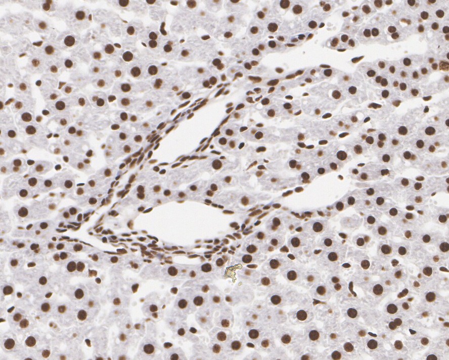
Immunohistochemical analysis of paraffin-embedded rat liver tissue with Rabbit anti-Phospho-JNK1/2/3 (T183 + T183 + T221) antibody at 1/1,000 dilution. The section was pre-treated using heat mediated antigen retrieval with sodium citrate buffer (pH 6.0) for 2 minutes. The tissues were blocked in 1% BSA for 20 minutes at room temperature, washed with ddH2O and PBS, and then probed with the primary antibody at 1/1,000 dilution for 1 hour at room temperature. The detection was performed using an HRP conjugated compact polymer system. DAB was used as the chromogen. Tissues were counterstained with hematoxylin and mounted with DPX.ICC/IF
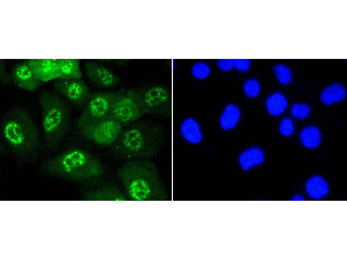
ICC staining of Phospho-JNK1/2/3 (T183 + T183 + T221) in HUVEC cells (green). Formalin fixed cells were permeabilized with 0.1% Triton X-100 in TBS for 10 minutes at room temperature and blocked with 1% Blocker BSA for 15 minutes at room temperature. Cells were probed with the primary antibody (1/50) for 1 hour at room temperature, washed with PBS. Alexa Fluor®488 Goat anti-Rabbit IgG was used as the secondary antibody at 1/1,000 dilution. The nuclear counter stain is DAPI (blue).IF-F
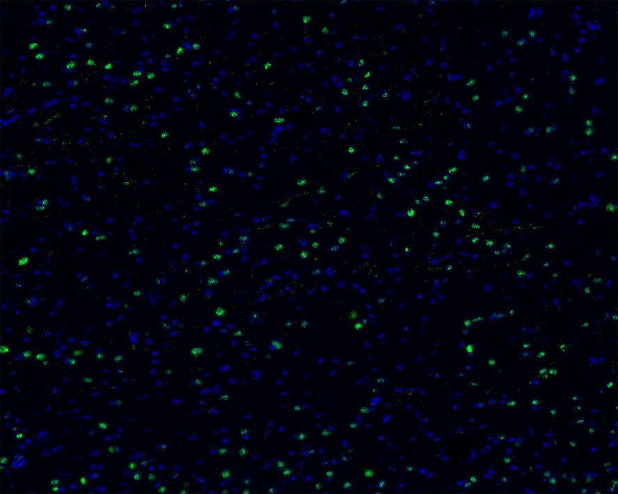
Immunofluorescence analysis of frozen mouse cerebral cortex tissue labeling Phospho-JNK1/2/3 (T183 + T183 + T221) with Rabbit anti-Phospho-JNK1/2/3 (T183 + T183 + T221) antibody. The tissues were blocked in 3% BSA for 30 minutes at room temperature, washed with PBS, and then probed with the primary antibody (green) at 1/100 dilution overnight at 4℃, washed with PBS. Goat Anti-Rabbit IgG H&L (Alexa Fluor® 488) was used as the secondary antibody at 1/200 dilution. Nuclei were counterstained with DAPI (blue). Image acquisition was performed with KFBIO KF-FL-400 Scanner.FC
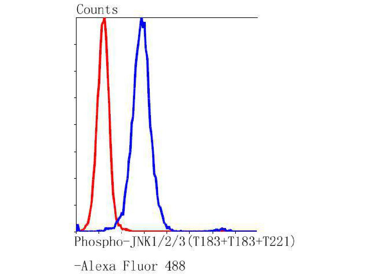
Flow cytometric analysis of Phospho-JNK1/2/3 (T183 + T183 + T221) was done on Hela cells. The cells were fixed, permeabilized and stained with the primary antibody (1/100) (blue). After incubation of the primary antibody at room temperature for an hour, the cells were stained with a Alexa Fluor 488-conjugated Goat anti-Rabbit IgG Secondary antibody at 1/1000 dilution for 30 minutes.Unlabelled sample was used as a control (cells without incubation with primary antibody; red).| Product Name | Phospho-JNK1/2/3 (T183 + T183 + T221) Recombinant Rabbit Monoclonal Antibody |
|---|---|
| Antibody Type | Primary Antibodies |
| Product description | JNKs (c-Jun N-terminal kinases) belong to a family of MAP kinases that are involved in a variety of cellular processes, including transcriptional regulation and cellular proliferation, differentiation and development. JNK2 (c-Jun N-terminal kinase 2) and JNK3 (c-Jun N-terminal kinase 3) are 424 and 464 amino acid proteins, respectively, that each contain one protein kinase domain and use magnesium as a cofactor to catalyze the phosphorylation of target proteins, thereby playing a role in a variety of events throughout the cell. Both JNK2 and JNK3 exist as multiple alternatively spliced isoforms and are subject to post-translational phosphorylation on Thr 183 and Thr 221, respectively, an event which activates JNK2/JNK3 enzymatic activity. Defects in the gene encoding JNK3 are a cause of epileptic encephalopathy of the Lennox-Gastaut type, a group of epileptic disorders characterized by severe psychomotor delay and seizures. |
| Immunogen | Synthetic phospho-peptide corresponding to residues surrounding Thr183 of Human JNK1 aa 161-204 / 427. |
| Modification | p-T183+T183+T221 |
| Clonality | Monoclonal |
|---|---|
| Isotype | IgG |
| Host Species | Recombinant rabbit |
| Tested Applications | FCICC/IFIF-FIHCWB |
| WB:1:2000-1:5000 IHC:1:400-1:1000 ICC/IF:1:100-1:200 IF-F:1:100-1:500 FC:1:500-1:1000 |
|
| Species Reactivity | HumanMouseRat |
| Concentration | 1mg/ml |
| Purification | Protein A |
| Alternative Names | C Jun kinase 2 antibody c Jun N terminal kinase 1 antibody c Jun N terminal kinase 2 antibody c Jun N terminal kinase 3 antibody c-Jun N-terminal kinase 1 antibody JNK 46 antibody JNK 55 antibody JNK antibody JNK-46 antibody JNK1 antibody JNK1A2 antibody JNK2 antibody JNK21B1/2 antibody JNK2A antibody JNK2ALPHA antibody JNK2B antibody JNK2BETA antibody JNK3 alpha protein kinase antibody JNK3 antibody JNK3A antibody Jun kinase antibody JUN N terminal kinase antibody MAP kinase 10 antibody MAP kinase 8 antibody MAP kinase 9 antibody MAP kinase p49 3F12 antibody MAPK 10 antibody MAPK 8 antibody MAPK 9 antibody MAPK10 antibody mapk8 antibody MAPK9 antibody Mitogen activated protein kinase 10 antibody Mitogen activated protein kinase 8 antibody Mitogen activated protein kinase 8 isoform JNK1 alpha1 antibody Mitogen activated protein kinase 8 isoform JNK1 beta2 antibody Mitogen activated protein kinase 9 antibody Mitogen-activated protein kinase 8 antibody MK08_HUMAN antibody p493F12 antibody p54a antibody p54aSAPK antibody p54bSAPK antibody PRKM10 antibody PRKM8 antibody PRKM9 antibody SAPK antibody SAPK(beta) antibody SAPK1 antibody SAPK1a antibody SAPK1b antibody SAPK1c antibody Stress activated protein kinase 1 antibody Stress activated protein kinase 1a antibody Stress activated protein kinase 1b antibody Stress activated protein kinase 1c antibody Stress activated protein kinase beta antibody Stress activated protein kinase JNK1 antibody Stress activated protein kinase JNK2 antibody Stress activated protein kinase JNK3 antibody Stress-activated protein kinase 1c antibody Stress-activated protein kinase JNK1 antibody |
|---|---|
| Molecular Weight(MW) | 48/53 kDa |
| Cellular Localization | Cytoplasm, Membrane, Mitochondrion, Nucleus. |
WB
Western blot analysis of Phospho-JNK1/2/3 (T183 + T183 + T221) on different lysates with Rabbit anti-Phospho-JNK1/2/3 (T183 + T183 + T221) antibody at 1/2,000 dilution. Lane 1: C6 cell lysate, Lane 2: C6 treated with 25µg/mL Anisomycin for 30 minutes cell lysate. Lysates/proteins at 20 µg/Lane. Exposure time: 2 minutes 6 seconds; 4-20% SDS-PAGE gel. Proteins were transferred to a PVDF membrane and blocked with 5% NFDM/TBST for 1 hour at room temperature. The primary antibody at 1/2,000 dilution was used in 5% NFDM/TBST at 4℃ overnight. Goat Anti-Rabbit IgG - HRP Secondary Antibody at 1/50,000 dilution was used for 1 hour at room temperature.
WB
Western blot analysis of Phospho-JNK1/2/3 (T183 + T183 + T221) on different lysates with Rabbit anti-Phospho-JNK1/2/3 (T183 + T183 + T221) antibody at 1/2,000 dilution. Lane 1: HeLa cell lysate, Lane 2: HeLa treated with 25μg/mL Anisomycin for 30 minutes cell lysate, Lane 3: NIH/3T3 cell lysate, Lane 4: NIH/3T3 treated with 25μg/mL Anisomycin for 30 minutes cell lysate, Lysates/proteins at 20 µg/Lane. Exposure time: 59 seconds; 4-20% SDS-PAGE gel. Proteins were transferred to a PVDF membrane and blocked with 5% NFDM/TBST for 1 hour at room temperature. The primary antibody at 1/2,000 dilution was used in 5% NFDM/TBST at 4℃ overnight. Goat Anti-Rabbit IgG - HRP Secondary Antibody at 1/50,000 dilution was used for 1 hour at room temperature.
IHC
Immunohistochemical analysis of paraffin-embedded human colon tissue with Rabbit anti-Phospho-JNK1/2/3 (T183 + T183 + T221) antibody at 1/400 dilution. The section was pre-treated using heat mediated antigen retrieval with sodium citrate buffer (pH 6.0) for 2 minutes. The tissues were blocked in 1% BSA for 20 minutes at room temperature, washed with ddH2O and PBS, and then probed with the primary antibody at 1/400 dilution for 1 hour at room temperature. The detection was performed using an HRP conjugated compact polymer system. DAB was used as the chromogen. Tissues were counterstained with hematoxylin and mounted with DPX.
IHC
Immunohistochemical analysis of paraffin-embedded mouse heart tissue with Rabbit anti-Phospho-JNK1/2/3 (T183 + T183 + T221) antibody at 1/400 dilution. The section was pre-treated using heat mediated antigen retrieval with sodium citrate buffer (pH 6.0) for 2 minutes. The tissues were blocked in 1% BSA for 20 minutes at room temperature, washed with ddH2O and PBS, and then probed with the primary antibody at 1/400 dilution for 1 hour at room temperature. The detection was performed using an HRP conjugated compact polymer system. DAB was used as the chromogen. Tissues were counterstained with hematoxylin and mounted with DPX.
IHC
Immunohistochemical analysis of paraffin-embedded rat liver tissue with Rabbit anti-Phospho-JNK1/2/3 (T183 + T183 + T221) antibody at 1/1,000 dilution. The section was pre-treated using heat mediated antigen retrieval with sodium citrate buffer (pH 6.0) for 2 minutes. The tissues were blocked in 1% BSA for 20 minutes at room temperature, washed with ddH2O and PBS, and then probed with the primary antibody at 1/1,000 dilution for 1 hour at room temperature. The detection was performed using an HRP conjugated compact polymer system. DAB was used as the chromogen. Tissues were counterstained with hematoxylin and mounted with DPX.
ICC/IF
ICC staining of Phospho-JNK1/2/3 (T183 + T183 + T221) in HUVEC cells (green). Formalin fixed cells were permeabilized with 0.1% Triton X-100 in TBS for 10 minutes at room temperature and blocked with 1% Blocker BSA for 15 minutes at room temperature. Cells were probed with the primary antibody (1/50) for 1 hour at room temperature, washed with PBS. Alexa Fluor®488 Goat anti-Rabbit IgG was used as the secondary antibody at 1/1,000 dilution. The nuclear counter stain is DAPI (blue).
IF-F
Immunofluorescence analysis of frozen mouse cerebral cortex tissue labeling Phospho-JNK1/2/3 (T183 + T183 + T221) with Rabbit anti-Phospho-JNK1/2/3 (T183 + T183 + T221) antibody. The tissues were blocked in 3% BSA for 30 minutes at room temperature, washed with PBS, and then probed with the primary antibody (green) at 1/100 dilution overnight at 4℃, washed with PBS. Goat Anti-Rabbit IgG H&L (Alexa Fluor® 488) was used as the secondary antibody at 1/200 dilution. Nuclei were counterstained with DAPI (blue). Image acquisition was performed with KFBIO KF-FL-400 Scanner.
FC
Flow cytometric analysis of Phospho-JNK1/2/3 (T183 + T183 + T221) was done on Hela cells. The cells were fixed, permeabilized and stained with the primary antibody (1/100) (blue). After incubation of the primary antibody at room temperature for an hour, the cells were stained with a Alexa Fluor 488-conjugated Goat anti-Rabbit IgG Secondary antibody at 1/1000 dilution for 30 minutes.Unlabelled sample was used as a control (cells without incubation with primary antibody; red).| Application Notes | WB:1:2000-1:5000 IHC:1:400-1:1000 ICC/IF:1:100-1:200 IF-F:1:100-1:500 FC:1:500-1:1000 |
|---|
| Form | Liquid |
|---|---|
| Storage Instructions | Store at +4℃ after thawing. Aliquot store at -20℃ or -80℃. Avoid repeated freeze / thaw cycles. |
| Storage Buffer | 1*TBS (pH7.4), 1%BSA, 40%Glycerol. Preservative: 0.05% Sodium Azide. |


