WB
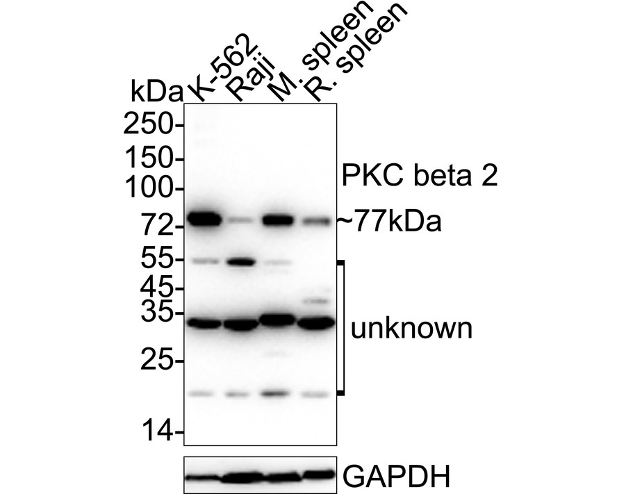
Western blot analysis of PKC beta 2 on different lysates with Rabbit anti-PKC beta 2 antibody at 1/500 dilution. Lane 1: K-562 cell lysate (20 µg/Lane), Lane 2: Raji cell lysate (20 µg/Lane), Lane 3: Mouse spleen tissue lysate (40 µg/Lane), Lane 4: Rat spleen tissue lysate (40 µg/Lane), Exposure time: 25 seconds; 4-20% SDS-PAGE gel. Proteins were transferred to a PVDF membrane and blocked with 5% NFDM/TBST for 1 hour at room temperature. The primary antibody at 1/500 dilution was used in 5% NFDM/TBST at 4℃ overnight. Goat Anti-Rabbit IgG - HRP Secondary Antibody at 1/50,000 dilution was used for 1 hour at room temperature.ICC/IF
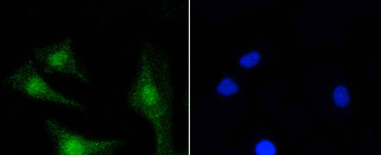
ICC staining PKC beta 2 in SH-SY-5Y cells (green). The nuclear counter stain is DAPI (blue). Cells were fixed in paraformaldehyde, permeabilised with 0.25% Triton X100/PBS.IHC
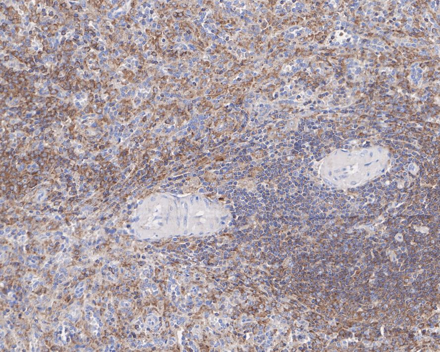
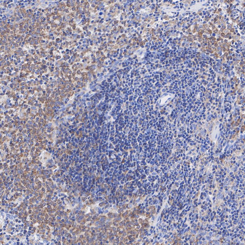
Immunohistochemical analysis of paraffin-embedded human spleen tissue with Rabbit anti-PKC beta 2 antibody at 1/50 dilution. The section was pre-treated using heat mediated antigen retrieval with sodium citrate buffer (pH 6.0) for 2 minutes. The tissues were blocked in 1% BSA for 20 minutes at room temperature, washed with ddH2O and PBS, and then probed with the primary antibody at 1/50 dilution for 1 hour at room temperature. The detection was performed using an HRP conjugated compact polymer system. DAB was used as the chromogen. Tissues were counterstained with hematoxylin and mounted with DPX.IHC
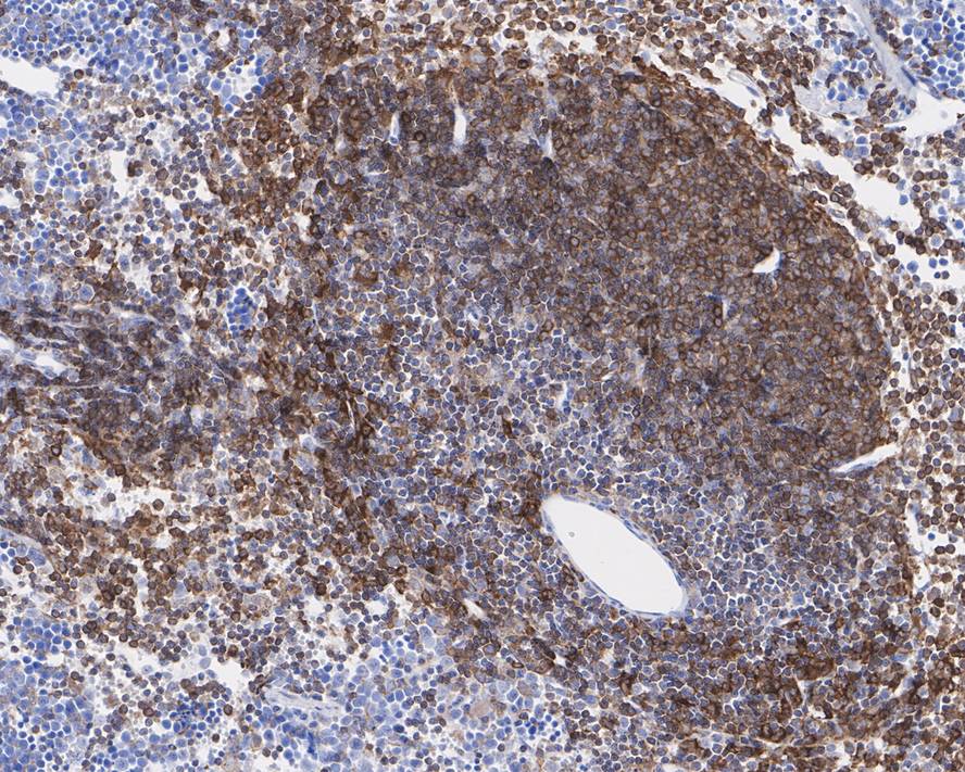
Immunohistochemical analysis of paraffin-embedded mouse spleen tissue with Rabbit anti-PKC beta 2 antibody at 1/200 dilution. The section was pre-treated using heat mediated antigen retrieval with sodium citrate buffer (pH 6.0) for 2 minutes. The tissues were blocked in 1% BSA for 20 minutes at room temperature, washed with ddH2O and PBS, and then probed with the primary antibody at 1/200 dilution for 1 hour at room temperature. The detection was performed using an HRP conjugated compact polymer system. DAB was used as the chromogen. Tissues were counterstained with hematoxylin and mounted with DPX.IHC
Immunohistochemical analysis of paraffin-embedded rat spleen tissue with Rabbit anti-PKC beta 2 antibody at 1/200 dilution. The section was pre-treated using heat mediated antigen retrieval with Tris-EDTA buffer (pH 9.0) for 20 minutes. The tissues were blocked in 1% BSA for 20 minutes at room temperature, washed with ddH2O and PBS, and then probed with the primary antibody at 1/200 dilution for 1 hour at room temperature. The detection was performed using an HRP conjugated compact polymer system. DAB was used as the chromogen. Tissues were counterstained with hematoxylin and mounted with DPX.FC
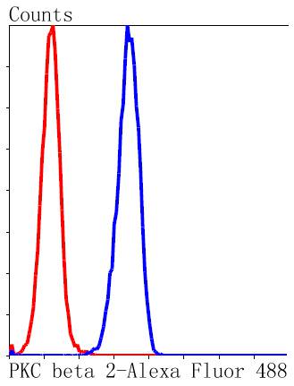
Flow cytometric analysis of Hela cells with PKC beta 2 antibody at 1/50 dilution (blue) compared with an unlabelled control (cells without incubation with primary antibody; red). Alexa Fluor 488-conjugated goat anti rabbit IgG was used as the secondary antibody.| Product Name | PKC beta 2 Recombinant Rabbit Monoclonal Antibody |
|---|---|
| Antibody Type | Primary Antibodies |
| Product description | Members of the protein kinase C (PKC) family play a key regulatory role in a variety of cellular functions including cell growth and differentiation, gene expression, hormone secretion and membrane function. PKCs were originally identified as serine/threonine protein kinases whose activity was dependent on calcium and phospholipids. Diacylglycerols (DAG) and tumor promoting phorbol esters bind to and activate PKC. PKCs can be subdivided into at least two major classes including conventional PKC isoforms (α, βI, βII and γ) and novel PKC isoforms. Patterns of expression for each PKC isoform differ among tissues and PKC family members exhibit clear differences in their cofactor dependencies. For instance, the kinase activities of nPKC δ and é are independent of Ca2+. On the other hand, nPKC δ and ε, as well as all of the cPKC members, possess phorbol ester-binding activities and kinase activities. |
| Immunogen | Synthetic peptide within Human PKC beta 2 aa 615-660 / 671. |
| Clonality | monoclonal |
|---|---|
| Isotype | IgG |
| Host Species | Recombinant rabbit |
| Tested Applications | FCICC/IFIHCWB |
| WB:1:500 IHC:1:50-1:200 ICC/IF:1:50-1:200 FC:1:50-1:100 |
|
| Species Reactivity | HumanMouseRat |
| Concentration | 1mg/ml |
| Purification | Protein A |
| Alternative Names | KPCB_HUMAN antibody PKC Beta antibody PKC-B antibody PKC-beta antibody PKCB antibody Prkcb antibody PRKCB II antibody PRKCB2 antibody Protein kinase C beta antibody Protein kinase C beta type antibody |
|---|---|
| Molecular Weight(MW) | 77kDa |
| Cellular Localization | Cytoplasm, Nucleus, Membrane. |
WB
Western blot analysis of PKC beta 2 on different lysates with Rabbit anti-PKC beta 2 antibody at 1/500 dilution. Lane 1: K-562 cell lysate (20 µg/Lane), Lane 2: Raji cell lysate (20 µg/Lane), Lane 3: Mouse spleen tissue lysate (40 µg/Lane), Lane 4: Rat spleen tissue lysate (40 µg/Lane), Exposure time: 25 seconds; 4-20% SDS-PAGE gel. Proteins were transferred to a PVDF membrane and blocked with 5% NFDM/TBST for 1 hour at room temperature. The primary antibody at 1/500 dilution was used in 5% NFDM/TBST at 4℃ overnight. Goat Anti-Rabbit IgG - HRP Secondary Antibody at 1/50,000 dilution was used for 1 hour at room temperature.
ICC/IF
ICC staining PKC beta 2 in SH-SY-5Y cells (green). The nuclear counter stain is DAPI (blue). Cells were fixed in paraformaldehyde, permeabilised with 0.25% Triton X100/PBS.
IHC
Immunohistochemical analysis of paraffin-embedded human spleen tissue with Rabbit anti-PKC beta 2 antibody at 1/50 dilution. The section was pre-treated using heat mediated antigen retrieval with sodium citrate buffer (pH 6.0) for 2 minutes. The tissues were blocked in 1% BSA for 20 minutes at room temperature, washed with ddH2O and PBS, and then probed with the primary antibody at 1/50 dilution for 1 hour at room temperature. The detection was performed using an HRP conjugated compact polymer system. DAB was used as the chromogen. Tissues were counterstained with hematoxylin and mounted with DPX.
IHC
Immunohistochemical analysis of paraffin-embedded mouse spleen tissue with Rabbit anti-PKC beta 2 antibody at 1/200 dilution. The section was pre-treated using heat mediated antigen retrieval with sodium citrate buffer (pH 6.0) for 2 minutes. The tissues were blocked in 1% BSA for 20 minutes at room temperature, washed with ddH2O and PBS, and then probed with the primary antibody at 1/200 dilution for 1 hour at room temperature. The detection was performed using an HRP conjugated compact polymer system. DAB was used as the chromogen. Tissues were counterstained with hematoxylin and mounted with DPX.
IHC
Immunohistochemical analysis of paraffin-embedded rat spleen tissue with Rabbit anti-PKC beta 2 antibody at 1/200 dilution. The section was pre-treated using heat mediated antigen retrieval with Tris-EDTA buffer (pH 9.0) for 20 minutes. The tissues were blocked in 1% BSA for 20 minutes at room temperature, washed with ddH2O and PBS, and then probed with the primary antibody at 1/200 dilution for 1 hour at room temperature. The detection was performed using an HRP conjugated compact polymer system. DAB was used as the chromogen. Tissues were counterstained with hematoxylin and mounted with DPX.
FC
Flow cytometric analysis of Hela cells with PKC beta 2 antibody at 1/50 dilution (blue) compared with an unlabelled control (cells without incubation with primary antibody; red). Alexa Fluor 488-conjugated goat anti rabbit IgG was used as the secondary antibody.| Application Notes | WB:1:500 IHC:1:50-1:200 ICC/IF:1:50-1:200 FC:1:50-1:100 |
|---|
| Form | Liquid |
|---|---|
| Storage Instructions | Store at +4℃ after thawing. Aliquot store at -20℃ or -80℃. Avoid repeated freeze / thaw cycles. |
| Storage Buffer | 1*TBS (pH7.4), 1%BSA, 40%Glycerol. Preservative: 0.05% Sodium Azide. |


