Application
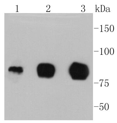
Fig1: Western blot analysis of ICAM1 on different lysates using anti-ICAM1 antibody at 1/1,000 dilution. Positive control: Lane 1: Hela Lane 2: HUVEC Lane 3: RajiApplication
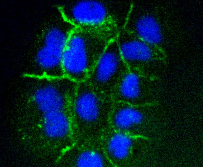
Fig2: ICC staining ICAM1 in A431 cells (green). The nuclear counter stain is DAPI (blue). Cells were fixed in paraformaldehyde, permeabilised with 0.25% Triton X100/PBS.Application
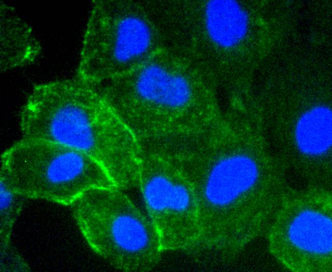
Fig3: ICC staining ICAM1 in HUVEC cells (green). The nuclear counter stain is DAPI (blue). Cells were fixed in paraformaldehyde, permeabilised with 0.25% Triton X100/PBS.Application
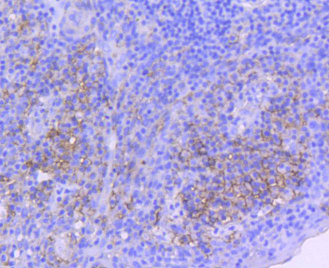
Fig4: Immunohistochemical analysis of paraffin-embedded human tonsil tissue using anti-ICAM1 antibody. Counter stained with hematoxylin.Application
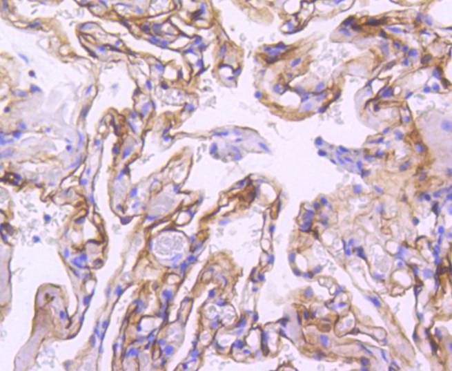
Fig5: Immunohistochemical analysis of paraffin-embedded human lung tissue using anti-ICAM1 antibody. Counter stained with hematoxylin.Application
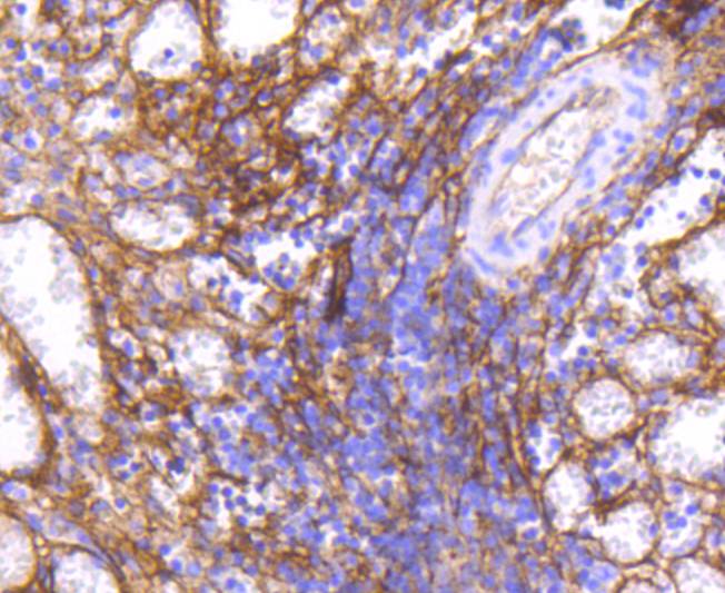
Fig6: Immunohistochemical analysis of paraffin-embedded human spleen tissue using anti-ICAM1 antibody. Counter stained with hematoxylin.Application
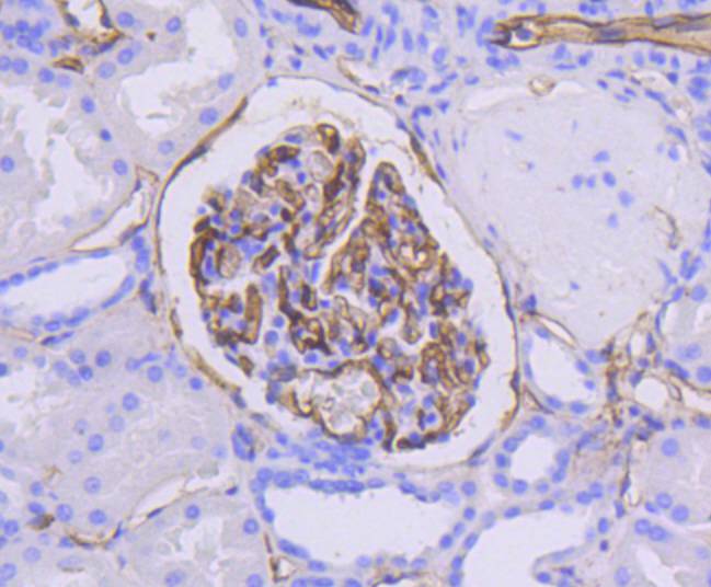
Fig7: Immunohistochemical analysis of paraffin-embedded human kidney tissue using anti-ICAM1 antibody. Counter stained with hematoxylin.| Product Name | ICAM1 [ST0487] |
|---|---|
| Antibody Type | Primary Antibodies |
| Antigen Alias | Antigen identified by monoclonal antibody BB2 antibody BB 2 antibody BB2 antibody CD 54 antibody CD_antigen=CD54 antibody CD54 antibody Cell surface glycoprotein P3.58 antibody Human rhinovirus receptor antibody ICAM 1 antibody ICAM-1 antibody ICAM1 antibody ICAM1_HUMAN antibody intercellular adhesion molecule 1 (CD54), human rhinovirus receptor antibody Intercellular adhesion molecule 1 antibody Major group rhinovirus receptor antibody MALA 2 antibody MALA2 antibody MyD 10 antibody MyD10 antibody P3.58 antibody Surface antigen of activated B cells, BB2 antibody |
| Product description | Cell adhesion molecules (CAMs) are a family of closely related cell surface glycoproteins involved in cell-cell interactions during growth and are thought to play important, yet separate, roles in embryogenesis and development. The intracellular adhesion molecule-1 (ICAM-1), also referred to as CD54, is an integral membrane protein of the immunoglobulin superfamily and recognizes the beta2alpha1 and beta2alphaM Integrins. ICAM-2 functions as a ligand for lymphocyte function-associated antigen-1 (LFA-1) and is involved in leukocyte adhesion. ICAM-3 is highly expressed on the surface of human eosinophils and, when bound to ligand, may inhibit eosinophil inflammatory responses and survival. ICAM-4, also known as LW glycoprotein, interacts with Integrins alphaLbeta2, alphaMbeta2, alpha4beta1, the alphaV family and alphaIIbbeta3, and selective binding to different integrins may be relevant to the pathology in a number of red blood cell associated diseases. Lastly, ICAM-5, expressed on telencephalic neurons, binds CD11a/CD18 and thus may act as an adhesion molecule for leukocyte binding in the central nervous system. |
| Immunogen | recombinant protein |
| Clonality | Monoclonal |
|---|---|
| Isotype | IgG |
| Host Species | Recombinant rabbit |
| Tested Applications | WBICC/IFIHC |
| WB:1:1,000-1:5,000 ICC:1:50-1:200 IHC:1:50-1:200 |
|
| Species Reactivity | Human |
| Concentration | 1mg/ml |
| Alternative Names | Antigen identified by monoclonal antibody
BB2 antibody
BB 2 antibody
BB2 antibody
CD 54 antibody
CD_antigen=CD54 antibody
CD54 antibody
Cell surface glycoprotein P3.58 antibody
Human rhinovirus receptor antibody
ICAM 1 antibody
ICAM-1 antibody
ICAM1 antibody
ICAM1_HUMAN antibody
intercellular adhesion molecule 1 (CD54) human rhinovirus receptor antibody Intercellular adhesion molecule 1 antibody Major group rhinovirus receptor antibody MALA 2 antibody MALA2 antibody MyD 10 antibody MyD10 antibody P3.58 antibody Surface antigen of activated B cells BB2 antibody |
|---|---|
| Molecular Weight(MW) | 89 kDa |
| Cellular Localization | Membrane. |
| SwissProt ID | P05362 |
|---|
Application
Fig1: Western blot analysis of ICAM1 on different lysates using anti-ICAM1 antibody at 1/1,000 dilution. Positive control: Lane 1: Hela Lane 2: HUVEC Lane 3: Raji
Application
Fig2: ICC staining ICAM1 in A431 cells (green). The nuclear counter stain is DAPI (blue). Cells were fixed in paraformaldehyde, permeabilised with 0.25% Triton X100/PBS.
Application
Fig3: ICC staining ICAM1 in HUVEC cells (green). The nuclear counter stain is DAPI (blue). Cells were fixed in paraformaldehyde, permeabilised with 0.25% Triton X100/PBS.
Application
Fig4: Immunohistochemical analysis of paraffin-embedded human tonsil tissue using anti-ICAM1 antibody. Counter stained with hematoxylin.
Application
Fig5: Immunohistochemical analysis of paraffin-embedded human lung tissue using anti-ICAM1 antibody. Counter stained with hematoxylin.
Application
Fig6: Immunohistochemical analysis of paraffin-embedded human spleen tissue using anti-ICAM1 antibody. Counter stained with hematoxylin.
Application
Fig7: Immunohistochemical analysis of paraffin-embedded human kidney tissue using anti-ICAM1 antibody. Counter stained with hematoxylin.| Positive Control | HUVEC, Raji, Hela, A431, human tonsil tissue, human spleen tissue, human lung tissue, human kidney tissue. |
|---|---|
| Application Notes | WB:1:1,000-1:5,000 ICC:1:50-1:200 IHC:1:50-1:200 |
| Form | Liquid |
|---|---|
| Storage Instructions | Store at +4℃ after thawing. Aliquot store at -20℃ or -80℃. Avoid repeated freeze / thaw cycles. |
| Storage Buffer | 1*TBS (pH7.4), 1%BSA, 40%Glycerol. Preservative: 0.05% Sodium Azide. |


