WB
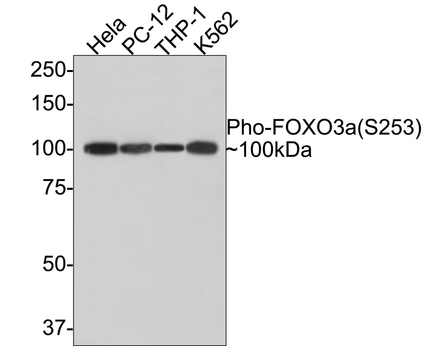
Western blot analysis of Phospho-FOXO3a (S253) on different lysates with Rabbit anti-Phospho-FOXO3a (S253) antibody at 1/1,000 dilution. Lane 1: Hela cell lysate, Lane 2: PC-12 cell lysate, Lane 3: THP-1 cell lysate, Lane 4: K562 cell lysate, Lysates/proteins at 10 µg/Lane. Exposure time: 1 minute; 8% SDS-PAGE gel. Proteins were transferred to a PVDF membrane and blocked with 5% NFDM/TBST for 1 hour at room temperature. The primary antibody at 1/1,000 dilution was used in 5% NFDM/TBST at room temperature for 2 hours. Goat Anti-Rabbit IgG - HRP Secondary Antibody at 1:300,000 dilution was used for 1 hour at room temperature.IHC
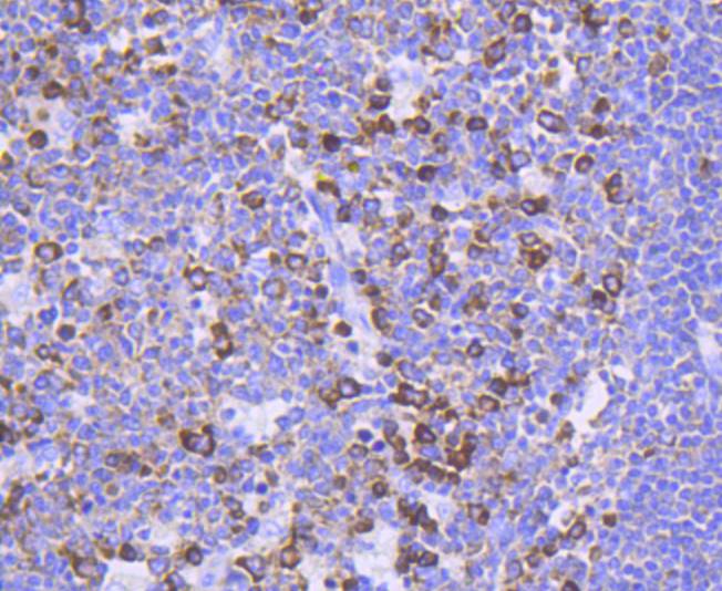
Immunohistochemical analysis of paraffin-embedded human tonsil tissue using anti-Phospho-FOXO3a(S253) antibody. Counter stained with hematoxylin.IHC
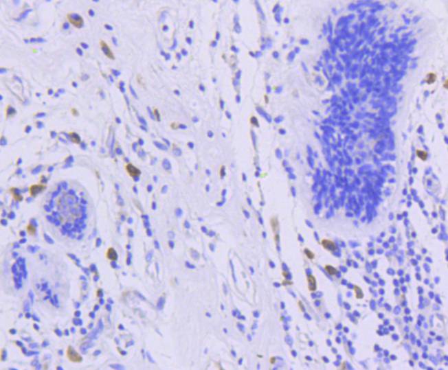
Immunohistochemical analysis of paraffin-embedded human breast carcinoma tissue using anti-Phospho-FOXO3a(S253) antibody. Counter stained with hematoxylin.IHC
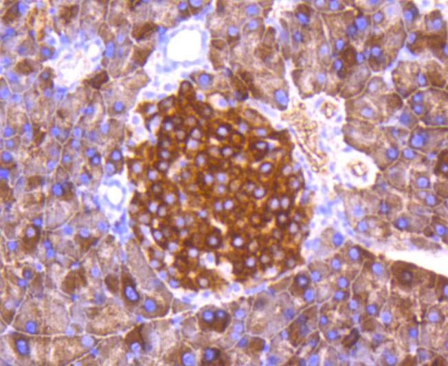
Immunohistochemical analysis of paraffin-embedded human pancreas tissue using anti-Phospho-FOXO3a(S253) antibody. Counter stained with hematoxylin.IHC
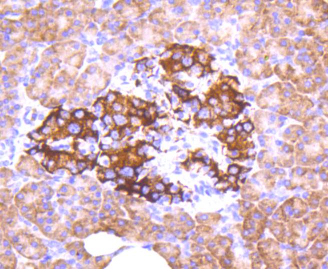
Immunohistochemical analysis of paraffin-embedded mouse pancreas tissue using anti-Phospho-FOXO3a(S253) antibody. Counter stained with hematoxylin.ICC/IF
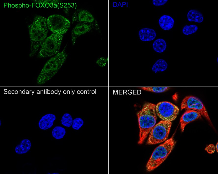
Immunocytochemistry analysis of NIH/3T3 cells labeling Phospho-FOXO3a (S253) with Rabbit anti-Phospho-FOXO3a (S253) antibody at 1/100 dilution. Cells were fixed in 4% paraformaldehyde for 15 minutes at room temperature, permeabilized with 0.1% Triton X-100 in PBS for 15 minutes at room temperature, then blocked with 1% BSA in 10% negative goat serum for 1 hour at room temperature. Cells were then incubated with Rabbit anti-Phospho-FOXO3a (S253) antibody at 1/100 dilution in 1% BSA in PBST overnight at 4 ℃. Goat Anti-Rabbit IgG H&L (488) was used as the secondary antibody at 1/1,000 dilution. PBS instead of the primary antibody was used as the secondary antibody only control. Nuclear DNA was labelled in blue with DAPI. Beta tubulin (red) was stained at 1/100 dilution overnight at +4℃. Goat Anti-Mouse IgG H&L (594) was used as the secondary antibody at 1/1,000 dilution.FC
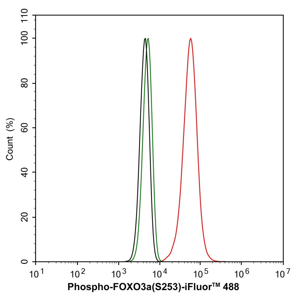
Flow cytometric analysis of NIH/3T3 cells labeling Phospho-FOXO3a (S253). Cells were fixed and permeabilized. Then stained with the primary antibody (1μg/mL) (red) compared with Rabbit IgG Isotype Control (green). After incubation of the primary antibody at +4℃ for an hour, the cells were stained with a 488 conjugate-Goat anti-Rabbit IgG Secondary antibody at 1/1,000 dilution for 30 minutes at +4℃. Unlabelled sample was used as a control (cells without incubation with primary antibody; black).| Product Name | Phospho-FOXO3a (S253) Recombinant Rabbit Monoclonal Antibody |
|---|---|
| Antibody Type | Primary Antibodies |
| Product description | FKHRL1 (for forkhead in rhabdomyosarcoma) is a member of the FKHR subfamily of forkhead transcription factors. Transcriptional activation of FKHR proteins is regulated by the serine/threonine kinase Akt1, which phosphorylates FKHRL1 at threonine-32 and serine-253. Phosphorylation by Akt1 negatively regulates FKHRL1 by promoting its export from the nucleus. Phosphorylated FKHRL1 associates with 14-3-3 proteins and this complex is retained in the cytoplasm. Growth factor withdrawal stimulates FKHRL1 dephosphorylation and nuclear translocation, leading to FKHR-induced gene-specific transcriptional activation. Within the nucleus, dephospho-rylated FKHRL1 triggers apoptosis by inducing the expression of genes that are critical for cell death. |
| Immunogen | Synthetic phospho-peptide corresponding to residues surrounding Ser253 of human FOXO3a. |
| Modification | p-S253 |
| Clonality | Monoclonal |
|---|---|
| Isotype | IgG |
| Host Species | Recombinant rabbit |
| Tested Applications | FCICC/IFIHCWB |
| WB:1:1000-1:5000 IHC:1:50-1:200 ICC/IF:1:100 FC:1:1000 |
|
| Species Reactivity | HumanMouseRat |
| Concentration | 1mg/ml |
| Purification | Protein A |
| Alternative Names | AF6q21 antibody AF6q21 protein antibody DKFZp781A0677 antibody FKHR2 antibody FKHRL 1 antibody FKHRL1 antibody FKHRL1P2 antibody Forkhead (Drosophila) homolog (rhabdomyosarcoma) like 1 antibody Forkhead box O3 antibody Forkhead box O3A antibody Forkhead box protein O3 antibody Forkhead box protein O3A antibody Forkhead Drosophila homolog of in rhabdomyosarcoma like 1 antibody Forkhead homolog (rhabdomyosarcoma) like 1 antibody Forkhead in rhabdomyosarcoma like 1 antibody Forkhead in rhabdomyosarcoma-like 1 antibody FOX O3A antibody FOXO2 antibody foxo3 antibody FOXO3_HUMAN antibody FOXO3A antibody MGC12739 antibody MGC31925 antibody |
|---|---|
| Molecular Weight(MW) | 71 kDa(Observed band size: 100 kDa) |
| Cellular Localization | Cytoplasm, Nucleus. |
WB
Western blot analysis of Phospho-FOXO3a (S253) on different lysates with Rabbit anti-Phospho-FOXO3a (S253) antibody at 1/1,000 dilution. Lane 1: Hela cell lysate, Lane 2: PC-12 cell lysate, Lane 3: THP-1 cell lysate, Lane 4: K562 cell lysate, Lysates/proteins at 10 µg/Lane. Exposure time: 1 minute; 8% SDS-PAGE gel. Proteins were transferred to a PVDF membrane and blocked with 5% NFDM/TBST for 1 hour at room temperature. The primary antibody at 1/1,000 dilution was used in 5% NFDM/TBST at room temperature for 2 hours. Goat Anti-Rabbit IgG - HRP Secondary Antibody at 1:300,000 dilution was used for 1 hour at room temperature.
IHC
Immunohistochemical analysis of paraffin-embedded human tonsil tissue using anti-Phospho-FOXO3a(S253) antibody. Counter stained with hematoxylin.
IHC
Immunohistochemical analysis of paraffin-embedded human breast carcinoma tissue using anti-Phospho-FOXO3a(S253) antibody. Counter stained with hematoxylin.
IHC
Immunohistochemical analysis of paraffin-embedded human pancreas tissue using anti-Phospho-FOXO3a(S253) antibody. Counter stained with hematoxylin.
IHC
Immunohistochemical analysis of paraffin-embedded mouse pancreas tissue using anti-Phospho-FOXO3a(S253) antibody. Counter stained with hematoxylin.
ICC/IF
Immunocytochemistry analysis of NIH/3T3 cells labeling Phospho-FOXO3a (S253) with Rabbit anti-Phospho-FOXO3a (S253) antibody at 1/100 dilution. Cells were fixed in 4% paraformaldehyde for 15 minutes at room temperature, permeabilized with 0.1% Triton X-100 in PBS for 15 minutes at room temperature, then blocked with 1% BSA in 10% negative goat serum for 1 hour at room temperature. Cells were then incubated with Rabbit anti-Phospho-FOXO3a (S253) antibody at 1/100 dilution in 1% BSA in PBST overnight at 4 ℃. Goat Anti-Rabbit IgG H&L (488) was used as the secondary antibody at 1/1,000 dilution. PBS instead of the primary antibody was used as the secondary antibody only control. Nuclear DNA was labelled in blue with DAPI. Beta tubulin (red) was stained at 1/100 dilution overnight at +4℃. Goat Anti-Mouse IgG H&L (594) was used as the secondary antibody at 1/1,000 dilution.
FC
Flow cytometric analysis of NIH/3T3 cells labeling Phospho-FOXO3a (S253). Cells were fixed and permeabilized. Then stained with the primary antibody (1μg/mL) (red) compared with Rabbit IgG Isotype Control (green). After incubation of the primary antibody at +4℃ for an hour, the cells were stained with a 488 conjugate-Goat anti-Rabbit IgG Secondary antibody at 1/1,000 dilution for 30 minutes at +4℃. Unlabelled sample was used as a control (cells without incubation with primary antibody; black).| Application Notes | WB:1:1000-1:5000 IHC:1:50-1:200 ICC/IF:1:100 FC:1:1000 |
|---|
| Form | Liquid |
|---|---|
| Storage Instructions | Store at +4℃ after thawing. Aliquot store at -20℃ or -80℃. Avoid repeated freeze / thaw cycles. |
| Storage Buffer | 1*TBS (pH7.4), 1%BSA, 40%Glycerol. Preservative: 0.05% Sodium Azide. |


