WB
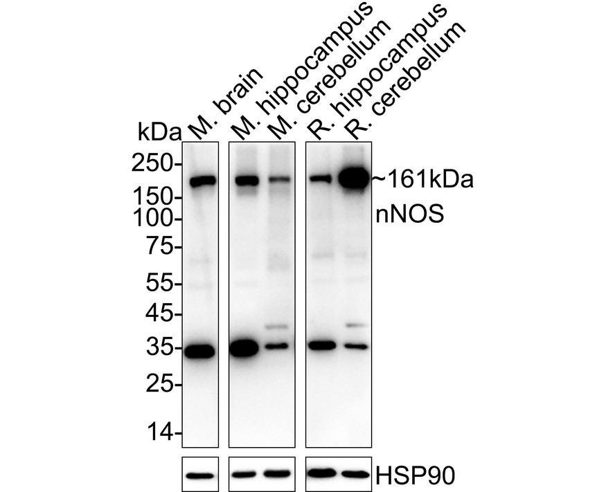
Western blot analysis of nNOS on different lysates with Rabbit anti-nNOS antibody at 1/1,000 dilution. Lane 1: Mouse brain tissue lysate (40 µg/Lane), Lane 2: Mouse hippocampus tissue lysate (40 µg/Lane), Lane 3: Mouse cerebellum tissue lysate (40 µg/Lane), Lane 4: Rat hippocampus tissue lysate (40 µg/Lane), Lane 5: Rat cerebellum tissue lysate (40 µg/Lane), Exposure time: 1 minute; 4-20% SDS-PAGE gel. Proteins were transferred to a PVDF membrane and blocked with 5% NFDM/TBST for 1 hour at room temperature. The primary antibody at 1/1,000 dilution was used in 5% NFDM/TBST at 4℃ overnight. Goat Anti-Rabbit IgG - HRP Secondary Antibody at 1/50,000 dilution was used for 1 hour at room temperature.IHC
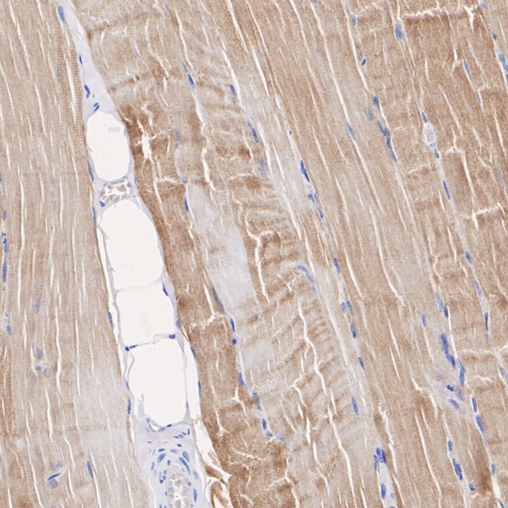
Immunohistochemical analysis of paraffin-embedded human skeletal muscle tissue with Rabbit anti-nNOS antibody at 1/200 dilution. The section was pre-treated using heat mediated antigen retrieval with Tris-EDTA buffer (pH 9.0) for 20 minutes. The tissues were blocked in 1% BSA for 20 minutes at room temperature, washed with ddH2O and PBS, and then probed with the primary antibody at 1/200 dilution for 1 hour at room temperature. The detection was performed using an HRP conjugated compact polymer system. DAB was used as the chromogen. Tissues were counterstained with hematoxylin and mounted with DPX.IHC
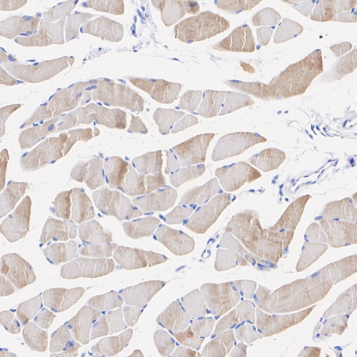
Immunohistochemical analysis of paraffin-embedded mouse skeletal muscle tissue with Rabbit anti-nNOS antibody at 1/200 dilution. The section was pre-treated using heat mediated antigen retrieval with Tris-EDTA buffer (pH 9.0) for 20 minutes. The tissues were blocked in 1% BSA for 20 minutes at room temperature, washed with ddH2O and PBS, and then probed with the primary antibody at 1/200 dilution for 1 hour at room temperature. The detection was performed using an HRP conjugated compact polymer system. DAB was used as the chromogen. Tissues were counterstained with hematoxylin and mounted with DPX.IHC
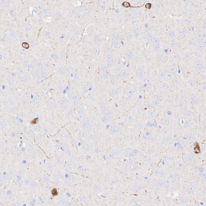
Immunohistochemical analysis of paraffin-embedded rat brain tissue with Rabbit anti-nNOS antibody at 1/1,000 dilution. The section was pre-treated using heat mediated antigen retrieval with Tris-EDTA buffer (pH 9.0) for 20 minutes. The tissues were blocked in 1% BSA for 20 minutes at room temperature, washed with ddH2O and PBS, and then probed with the primary antibody at 1/1,000 dilution for 1 hour at room temperature. The detection was performed using an HRP conjugated compact polymer system. DAB was used as the chromogen. Tissues were counterstained with hematoxylin and mounted with DPX.| Product Name | nNOS Recombinant Rabbit Monoclonal Antibody |
|---|---|
| Antibody Type | Primary Antibodies |
| Product description | Nitric oxide (NO) has a broad range of biological activities and has been implicated in signaling pathways in phylogenetically diverse species. Nitric oxide synthases (NOSs), the enzymes responsible for synthesis of NO, contain an N-terminal oxygenase domain and a C-terminal reductase domain. NOS activity requires homodimerization as well as three cosubstrates (L-arginine, NADPH and O2) and five cofactors or prosthetic groups (FAD, FMN, calmodulin, tetrahydrobiopterin and heme). Several distinct NOS isoforms have been described and been shown to represent the products of three distinct genes. These include two constitutive Ca2+/CaM-dependent forms of NOS, including NOS1 (also designated ncNOS) whose activity was first identified in neurons, and NOS3 (also designated ecNOS), first identified in endothelial cells. The inducible form of NOS, NOS2 (also designated iNOS), is Ca2+-independent and is expressed in a broad range of cell types. |
| Immunogen | Synthetic peptide within Human nNOS aa 1398-1434 / 1434. |
| Clonality | Monoclonal |
|---|---|
| Isotype | IgG |
| Host Species | Recombinant rabbit |
| Tested Applications | IHCWB |
| WB:1:1000-1:2000 IHC:1:200-1:1000 |
|
| Species Reactivity | HumanMouseRat |
| Concentration | 1mg/ml |
| Purification | Protein A |
| Alternative Names | 2310005C01Rik antibody
BNOS antibody
Constitutive NOS antibody
EC 1.14.13.39 antibody
IHPS 1 antibody
IHPS1 antibody
N-NOS antibody
NC-NOS antibody
neuronal Nitric Oxide Synthase antibody
Neuronal NOS antibody
Nitric oxide synthase neuronal included antibody Nitric oxide synthase 1 (neuronal) antibody Nitric oxide synthase 1 antibody Nitric oxide synthase brain antibody Nitric oxide synthase penile neuronal included antibody NNOS antibody NO antibody NOS 1 antibody NOS antibody NOS type I antibody NOS-I antibody NOS1 antibody NOS1_HUMAN antibody Peptidyl-cysteine S-nitrosylase NOS1 antibody |
|---|---|
| Molecular Weight(MW) | 161 kDa |
| Cellular Localization | Cell membrane, sarcolemma, Cell projection, dendritic spine. |
WB
Western blot analysis of nNOS on different lysates with Rabbit anti-nNOS antibody at 1/1,000 dilution. Lane 1: Mouse brain tissue lysate (40 µg/Lane), Lane 2: Mouse hippocampus tissue lysate (40 µg/Lane), Lane 3: Mouse cerebellum tissue lysate (40 µg/Lane), Lane 4: Rat hippocampus tissue lysate (40 µg/Lane), Lane 5: Rat cerebellum tissue lysate (40 µg/Lane), Exposure time: 1 minute; 4-20% SDS-PAGE gel. Proteins were transferred to a PVDF membrane and blocked with 5% NFDM/TBST for 1 hour at room temperature. The primary antibody at 1/1,000 dilution was used in 5% NFDM/TBST at 4℃ overnight. Goat Anti-Rabbit IgG - HRP Secondary Antibody at 1/50,000 dilution was used for 1 hour at room temperature.
IHC
Immunohistochemical analysis of paraffin-embedded human skeletal muscle tissue with Rabbit anti-nNOS antibody at 1/200 dilution. The section was pre-treated using heat mediated antigen retrieval with Tris-EDTA buffer (pH 9.0) for 20 minutes. The tissues were blocked in 1% BSA for 20 minutes at room temperature, washed with ddH2O and PBS, and then probed with the primary antibody at 1/200 dilution for 1 hour at room temperature. The detection was performed using an HRP conjugated compact polymer system. DAB was used as the chromogen. Tissues were counterstained with hematoxylin and mounted with DPX.
IHC
Immunohistochemical analysis of paraffin-embedded mouse skeletal muscle tissue with Rabbit anti-nNOS antibody at 1/200 dilution. The section was pre-treated using heat mediated antigen retrieval with Tris-EDTA buffer (pH 9.0) for 20 minutes. The tissues were blocked in 1% BSA for 20 minutes at room temperature, washed with ddH2O and PBS, and then probed with the primary antibody at 1/200 dilution for 1 hour at room temperature. The detection was performed using an HRP conjugated compact polymer system. DAB was used as the chromogen. Tissues were counterstained with hematoxylin and mounted with DPX.
IHC
Immunohistochemical analysis of paraffin-embedded rat brain tissue with Rabbit anti-nNOS antibody at 1/1,000 dilution. The section was pre-treated using heat mediated antigen retrieval with Tris-EDTA buffer (pH 9.0) for 20 minutes. The tissues were blocked in 1% BSA for 20 minutes at room temperature, washed with ddH2O and PBS, and then probed with the primary antibody at 1/1,000 dilution for 1 hour at room temperature. The detection was performed using an HRP conjugated compact polymer system. DAB was used as the chromogen. Tissues were counterstained with hematoxylin and mounted with DPX.| Application Notes | WB:1:1000-1:2000 IHC:1:200-1:1000 |
|---|
| Form | Liquid |
|---|---|
| Storage Instructions | Store at +4℃ after thawing. Aliquot store at -20℃ or -80℃. Avoid repeated freeze / thaw cycles. |
| Storage Buffer | 1*TBS (pH7.4), 1%BSA, 40%Glycerol. Preservative: 0.05% Sodium Azide. |


