WB
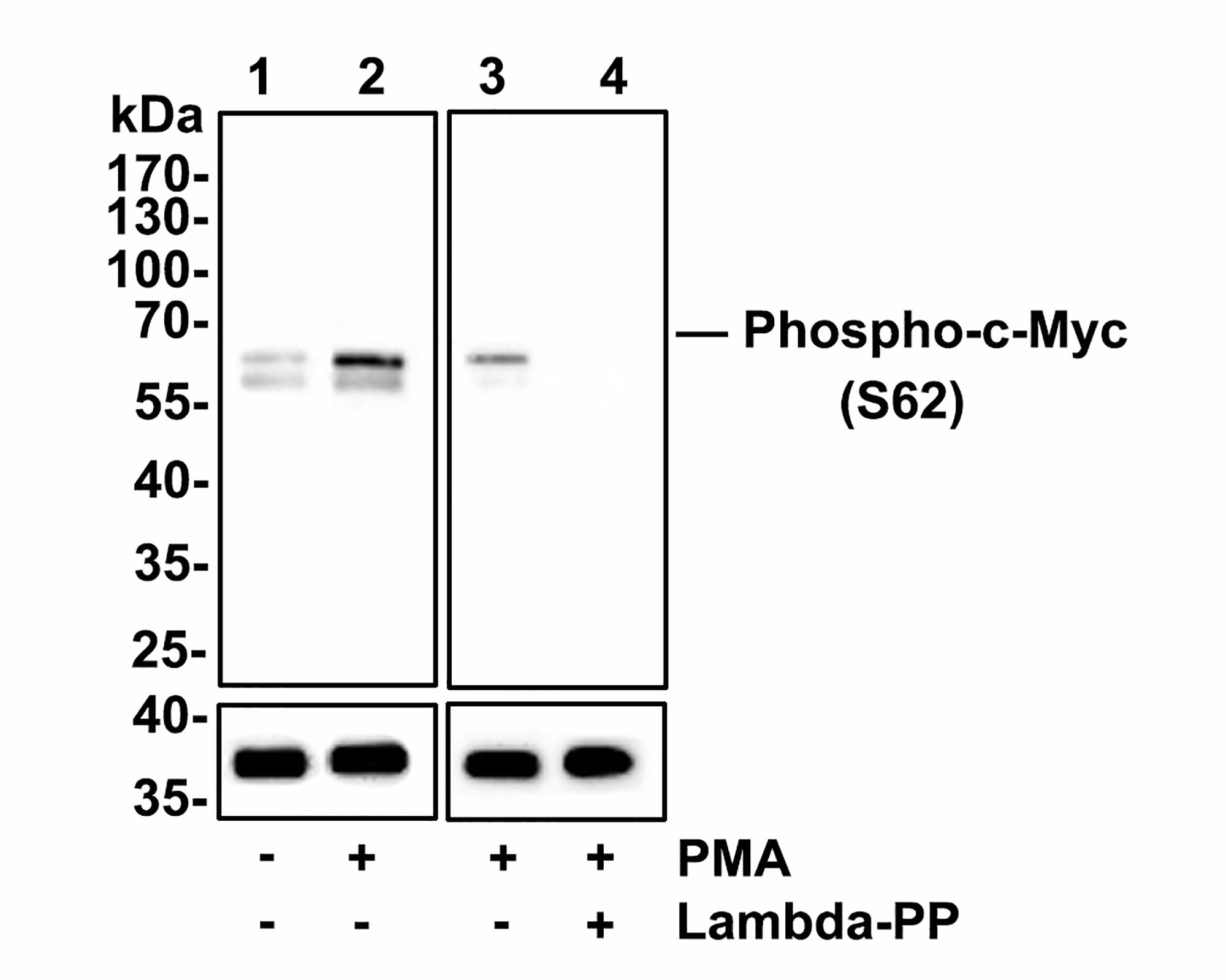
Western blot analysis of Phospho-c-Myc(S62) on HeLa cell lysates. Lane 1: HeLa cells, whole cell lysate, 10ug/lane, Lane 2/3: HeLa cells treated with 200 nM PMA for 10 minutes, whole cell lysates, 10ug/lane, Lane 4: HeLa cells treated with 200 nM PMA for 10 minutes, then treated with 2.8ug/ul lambda-PP for 30 minutes, whole cell lysates, 10ug/lane, All lanes : Anti-Phospho-c-Myc(S62) antibody at 1:500 dilution. Anti-GAPDH antibody at 1:10,000 dilution. Goat Anti-Rabbit IgG H&L (HRP)at 1/200,000 dilution. Blocking and diluting buffer: 5% BSA. Exposure time: 3 minutes.WB
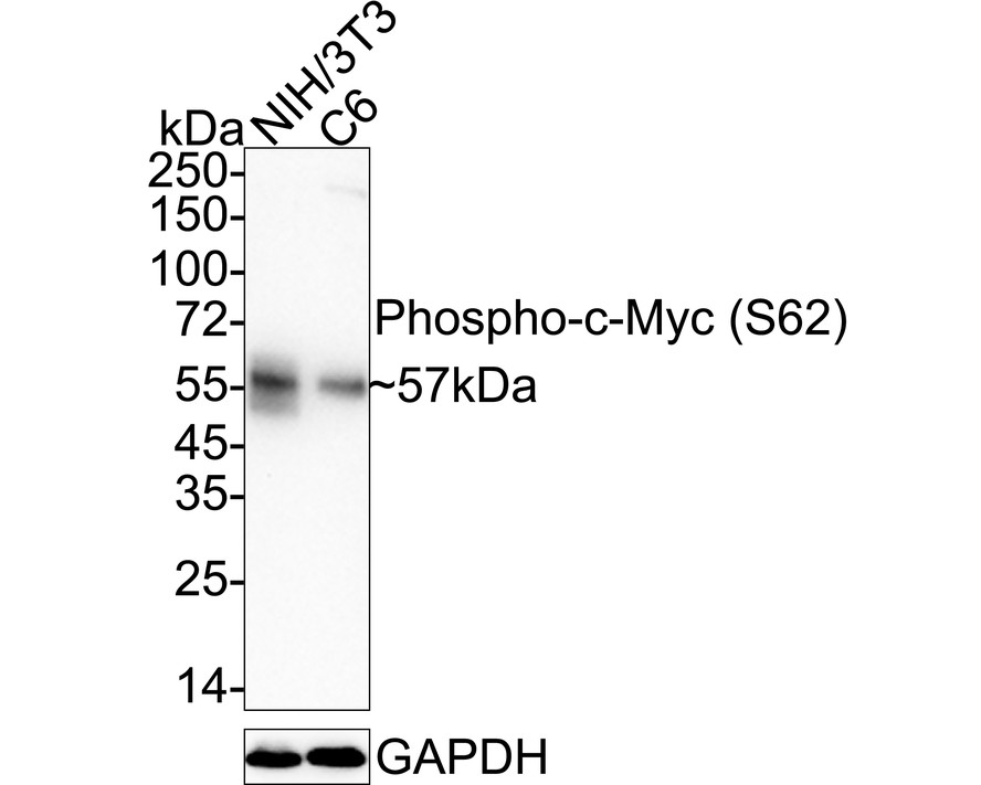
Western blot analysis of Phospho-c-Myc (S62) on different lysates with Rabbit anti-Phospho-c-Myc (S62) antibody at 1/1,000 dilution. Lane 1: NIH/3T3 cell lysate, Lane 2: C6 cell lysate, Lysates/proteins at 20 µg/Lane. Exposure time: 3 minutes; 4-20% SDS-PAGE gel. Proteins were transferred to a PVDF membrane and blocked with 5% NFDM/TBST for 1 hour at room temperature. The primary antibody at 1/1,000 dilution was used in 5% NFDM/TBST at 4℃ overnight. Goat Anti-Rabbit IgG - HRP Secondary Antibody at 1/50,000 dilution was used for 1 hour at room temperature.IHC
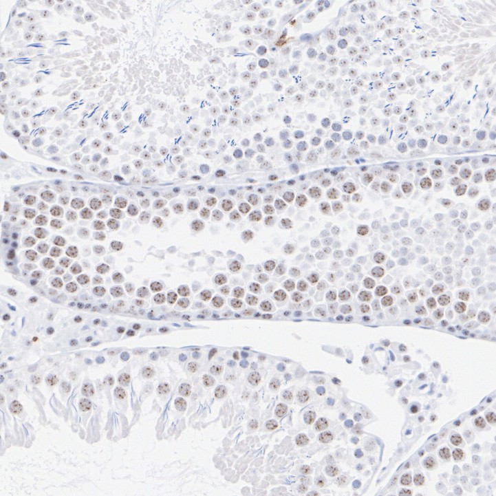
Immunohistochemical analysis of paraffin-embedded rat testis tissue with Rabbit anti-Phospho-c-Myc (S62) antibody at 1/200 dilution. The section was pre-treated using heat mediated antigen retrieval with sodium citrate buffer (pH 6.0) (high pressure) for 2 minutes. The tissues were blocked in 1% BSA for 20 minutes at room temperature, washed with ddH2O and PBS, and then probed with the primary antibody at 1/200 dilution for 1 hour at room temperature. The detection was performed using an HRP conjugated compact polymer system. DAB was used as the chromogen. Tissues were counterstained with hematoxylin and mounted with DPX.ICC/IF
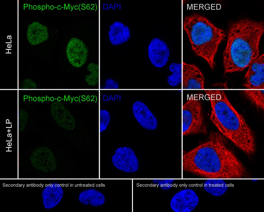
Immunocytochemistry analysis of HeLa cells treated with or without λpp labeling Phospho-c-Myc (S62) with Rabbit anti-Phospho-c-Myc (S62) antibody at 1/100 dilution. Cells were fixed in 4% paraformaldehyde for 15 minutes at room temperature, permeabilized with 0.1% Triton X-100 in PBS for 15 minutes at room temperature, then blocked with 1% BSA in 10% negative goat serum for 1 hour at room temperature. Cells were then incubated with Rabbit anti-Phospho-c-Myc (S62) antibody at 1/100 dilution in 1% BSA in PBST overnight at 4 ℃. Goat Anti-Rabbit IgG H&L (488) was used as the secondary antibody at 1/1,000 dilution. PBS instead of the primary antibody was used as the secondary antibody only control. Nuclear DNA was labelled in blue with DAPI. Beta tubulin (red) was stained at 1/100 dilution overnight at +4℃. Goat Anti-Mouse IgG H&L (594) was used as the secondary antibody at 1/1,000 dilution.| Product Name | Phospho-c-Myc (S62) Recombinant Rabbit Monoclonal Antibody |
|---|---|
| Antibody Type | Primary Antibodies |
| Product description | c-Myc-, N-Myc- and L-Myc-encoded proteins function in cell proliferation, differentiation and neoplastic disease. Myc proteins are nuclear proteins with relatively short half lives. Amplification of the c-Myc gene has been found in several types of human tumors including lung, breast and colon carcinomas, while the N-Myc gene has been found amplified in neuroblastomas. The L-Myc gene has been reported to be amplified and expressed at high level in human small cell lung carcinomas. The presence of three sequence motifs in the c-Myc COOH terminus, including the leucine zipper, the helix-loop-helix and a basic region provided initial evidence for a sequence-specific binding function. A basic region helix-loop-helix leucine zipper motif (bHLH-Zip) protein, designated Max, specifically associates with c-Myc, N-Myc and L-Myc proteins. The Myc-Max complex binds to DNA in a sequence-specific manner under conditions where neither Max nor Myc exhibit appreciable binding. Max can also form heterodimers with at least two additional bHLH-Zip proteins, Mad and Mxi1, and Mad-Max dimers have been shown to repress transcription through interaction with mSin3. |
| Immunogen | Synthetic phospho-peptide corresponding to residues surrounding Ser62 of Human c-Myc aa 46-87 / 439. |
| Modification | p-S62 |
| Clonality | Monoclonal |
|---|---|
| Isotype | IgG |
| Host Species | Recombinant rabbit |
| Tested Applications | ICC/IFIHCWB |
| WB:1:500-1:2000 IHC:1:50-1:200 ICC/IF:1:50-1:200 |
|
| Species Reactivity | HumanMouseRat |
| Concentration | 1mg/ml |
| Purification | Protein A |
| Alternative Names | AU016757 antibody Avian myelocytomatosis viral oncogene homolog antibody bHLHe39 antibody c Myc antibody Class E basic helix-loop-helix protein 39 antibody MRTL antibody Myc antibody Myc protein antibody Myc proto oncogene protein antibody Myc proto-oncogene protein antibody myc-related translation/localization regulatory factor antibody MYC_HUMAN antibody Myc2 antibody MYCC antibody Myelocytomatosis oncogene antibody Niard antibody Nird antibody Oncogene Myc antibody OTTHUMP00000158589 antibody Proto-oncogene c-Myc antibody Protooncogene homologous to myelocytomatosis virus antibody RNCMYC antibody Transcription factor p64 antibody Transcriptional regulator Myc-A antibody V-Myc avian myelocytomatosis viral oncogene homolog antibody v-myc myelocytomatosis viral oncogene homolog (avian) antibody |
|---|---|
| Molecular Weight(MW) | 57 kDa |
| Cellular Localization | Nucleoplasm, nucleolus. |
WB
Western blot analysis of Phospho-c-Myc(S62) on HeLa cell lysates. Lane 1: HeLa cells, whole cell lysate, 10ug/lane, Lane 2/3: HeLa cells treated with 200 nM PMA for 10 minutes, whole cell lysates, 10ug/lane, Lane 4: HeLa cells treated with 200 nM PMA for 10 minutes, then treated with 2.8ug/ul lambda-PP for 30 minutes, whole cell lysates, 10ug/lane, All lanes : Anti-Phospho-c-Myc(S62) antibody at 1:500 dilution. Anti-GAPDH antibody at 1:10,000 dilution. Goat Anti-Rabbit IgG H&L (HRP)at 1/200,000 dilution. Blocking and diluting buffer: 5% BSA. Exposure time: 3 minutes.
WB
Western blot analysis of Phospho-c-Myc (S62) on different lysates with Rabbit anti-Phospho-c-Myc (S62) antibody at 1/1,000 dilution. Lane 1: NIH/3T3 cell lysate, Lane 2: C6 cell lysate, Lysates/proteins at 20 µg/Lane. Exposure time: 3 minutes; 4-20% SDS-PAGE gel. Proteins were transferred to a PVDF membrane and blocked with 5% NFDM/TBST for 1 hour at room temperature. The primary antibody at 1/1,000 dilution was used in 5% NFDM/TBST at 4℃ overnight. Goat Anti-Rabbit IgG - HRP Secondary Antibody at 1/50,000 dilution was used for 1 hour at room temperature.
IHC
Immunohistochemical analysis of paraffin-embedded rat testis tissue with Rabbit anti-Phospho-c-Myc (S62) antibody at 1/200 dilution. The section was pre-treated using heat mediated antigen retrieval with sodium citrate buffer (pH 6.0) (high pressure) for 2 minutes. The tissues were blocked in 1% BSA for 20 minutes at room temperature, washed with ddH2O and PBS, and then probed with the primary antibody at 1/200 dilution for 1 hour at room temperature. The detection was performed using an HRP conjugated compact polymer system. DAB was used as the chromogen. Tissues were counterstained with hematoxylin and mounted with DPX.
ICC/IF
Immunocytochemistry analysis of HeLa cells treated with or without λpp labeling Phospho-c-Myc (S62) with Rabbit anti-Phospho-c-Myc (S62) antibody at 1/100 dilution. Cells were fixed in 4% paraformaldehyde for 15 minutes at room temperature, permeabilized with 0.1% Triton X-100 in PBS for 15 minutes at room temperature, then blocked with 1% BSA in 10% negative goat serum for 1 hour at room temperature. Cells were then incubated with Rabbit anti-Phospho-c-Myc (S62) antibody at 1/100 dilution in 1% BSA in PBST overnight at 4 ℃. Goat Anti-Rabbit IgG H&L (488) was used as the secondary antibody at 1/1,000 dilution. PBS instead of the primary antibody was used as the secondary antibody only control. Nuclear DNA was labelled in blue with DAPI. Beta tubulin (red) was stained at 1/100 dilution overnight at +4℃. Goat Anti-Mouse IgG H&L (594) was used as the secondary antibody at 1/1,000 dilution.| Application Notes | WB:1:500-1:2000 IHC:1:50-1:200 ICC/IF:1:50-1:200 |
|---|
| Form | Liquid |
|---|---|
| Storage Instructions | Store at +4℃ after thawing. Aliquot store at -20℃ or -80℃. Avoid repeated freeze / thaw cycles. |
| Storage Buffer | 1*TBS (pH7.4), 1%BSA, 40%Glycerol. Preservative: 0.05% Sodium Azide. |


