Application
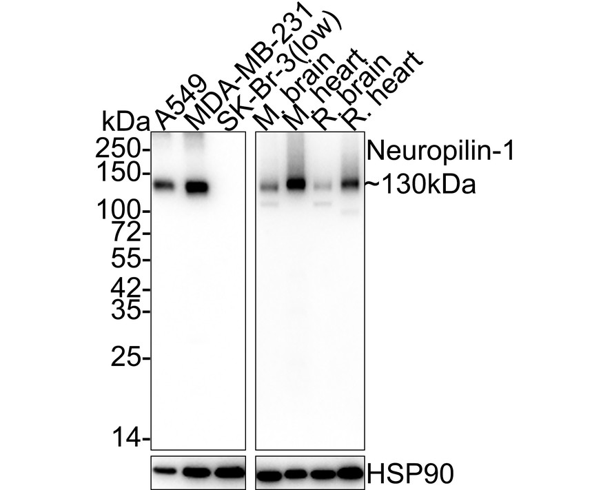
Fig1: Western blot analysis of Neuropilin 1 on different lysates with Rabbit anti-Neuropilin 1 antibody at 1/5,000 dilution. Lane 1: A549 cell lysate, Lane 2: MDA-MB-231 cell lysate, Lane 3: SK-Br-3 cell lysate (low expression), Lane 4: Mouse brain tissue lysate, Lane 5: Mouse heart tissue lysate, Lane 6: Rat brain tissue lysate, Lane 7: Rat heart tissue lysate, Lysates/proteins at 20 µg/Lane. Exposure time: Lane 1-3: 1 minute 21 seconds; Lane 4-7: 43 seconds; 4-20% SDS-PAGE gel. Proteins were transferred to a PVDF membrane and blocked with 5% NFDM/TBST for 1 hour at room temperature. The primary antibody at 1/5,000 dilution was used in 5% NFDM/TBST at room temperature for 2 hours. Goat Anti-Rabbit IgG - HRP Secondary Antibody at 1:100,000 dilution was used for 1 hour at room temperature.Application
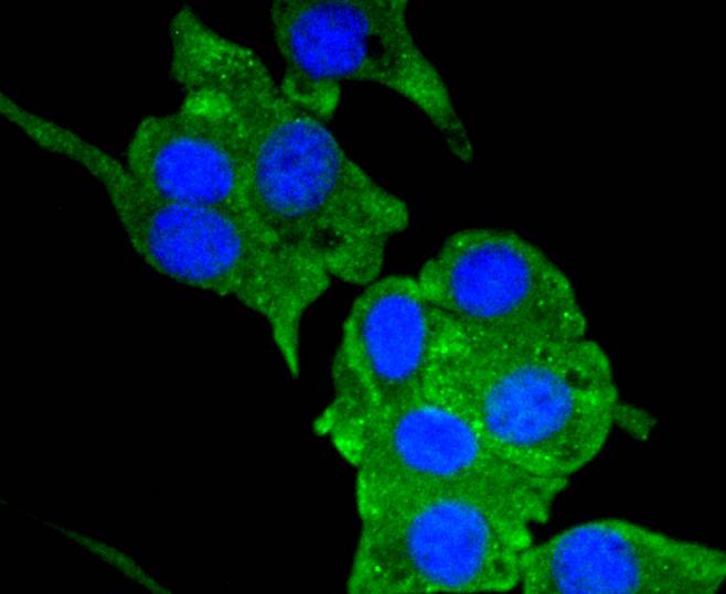
Fig2: ICC staining Neuropilin-1 in SHG-44 cells (green). The nuclear counter stain is DAPI (blue). Cells were fixed in paraformaldehyde, permeabilised with 0.25% Triton X100/PBS.Application
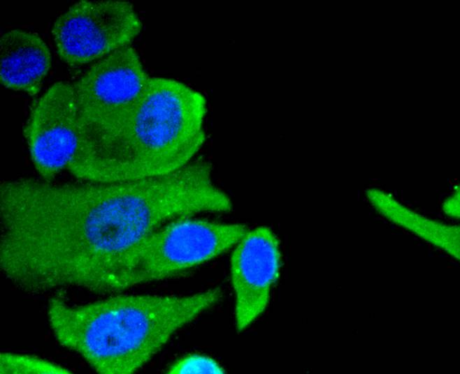
Fig3: ICC staining Neuropilin-1 in MCF-7 cells (green). The nuclear counter stain is DAPI (blue). Cells were fixed in paraformaldehyde, permeabilised with 0.25% Triton X100/PBS.Application
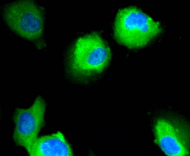
Fig4: ICC staining Neuropilin-1 in HUVEC cells (green). The nuclear counter stain is DAPI (blue). Cells were fixed in paraformaldehyde, permeabilised with 0.25% Triton X100/PBS.Application
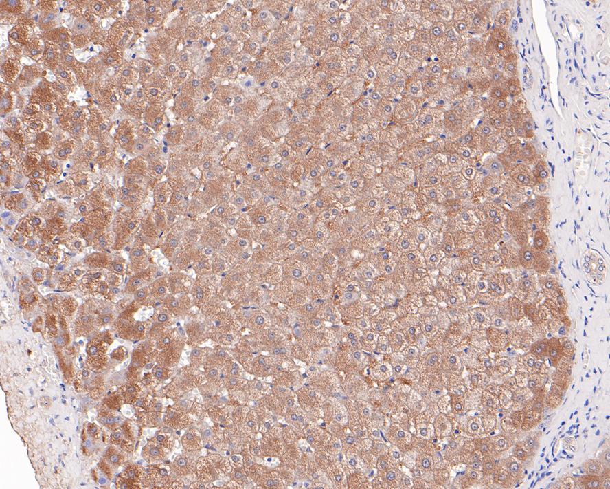
Fig5: Immunohistochemical analysis of paraffin-embedded human liver tissue with Rabbit anti-Neuropilin 1 antibody at 1/200 dilution. The section was pre-treated using heat mediated antigen retrieval with Tris-EDTA buffer (pH 9.0) for 20 minutes. The tissues were blocked in 1% BSA for 20 minutes at room temperature, washed with ddH2O and PBS, and then probed with the primary antibody at 1/200 dilution for 1 hour at room temperature. The detection was performed using an HRP conjugated compact polymer system. DAB was used as the chromogen. Tissues were counterstained with hematoxylin and mounted with DPX.Application
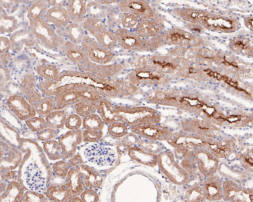
Fig6:Immunohistochemical analysis of paraffin-embedded mouse kidney tissue with Rabbit anti-Neuropilin 1 antibody at 1/200 dilution. The section was pre-treated using heat mediated antigen retrieval with Tris-EDTA buffer (pH 9.0) for 20 minutes. The tissues were blocked in 1% BSA for 20 minutes at room temperature, washed with ddH2O and PBS, and then probed with the primary antibody at 1/200 dilution for 1 hour at room temperature. The detection was performed using an HRP conjugated compact polymer system. DAB was used as the chromogen. Tissues were counterstained with hematoxylin and mounted with DPX.| Product Name | Neuropilin 1 Recombinant Rabbit Monoclonal Antibody |
|---|---|
| Antibody Type | Primary Antibodies |
| Product description | Neuropilin is a type I transmembrane receptor that has been implicated in aspects of axon growth and guidance and has been shown to act as a high affinity receptor for class III semaphorins and vascular endothelial growth factor (VEGF). A closely related protein, neuropilin-2, shares a common domain structure and signifcant homology with neuropilin and also acts as a receptor for the class III semaphorins and VEGF. Both neuropilins are involved in regulating many physiological pathways including axonal guidance and angiogenesis, however they exhibit differential expression in the adult vasculature. Neuropilin-2 is polysialylated and expressed on the surface of dendritic cells. It is also expressed by venous and lymphatic endothelium. Neuropilin is expressed predominantly by arterial endothelium. |
| Immunogen | Synthetic peptide within Human Neuropilin-1 aa 881-923 / 923. |
| Clonality | monoclonal |
|---|---|
| Isotype | IgG |
| Host Species | Recombinant rabbit |
| Tested Applications | ICC/IFIHCWB |
| WB:1:2000-1:5000 ICC/IF:1:50-1:200 IHC:1:50-1:200 |
|
| Species Reactivity | HumanMouseRat |
| Concentration | 1mg/ml |
| Purification | Protein A |
| Alternative Names | A5 protein antibody BDCA4 antibody BLOOD DENDRITIC CELL ANTIGEN 4 antibody CD304 antibody Neuropilin-1 antibody Neuropilin1 antibody NP1 antibody NPN1 antibody NRP 1 antibody NRP antibody NRP1 antibody NRP1_HUMAN antibody transmembrane receptor antibody Vascular endothelial cell growth factor 165 receptor antibody VEGF165R antibody |
|---|---|
| Molecular Weight(MW) | 103kDa(Observed band size: 130kDa) |
| Cellular Localization | Cell membrane, Secreted. |
Application
Fig1: Western blot analysis of Neuropilin 1 on different lysates with Rabbit anti-Neuropilin 1 antibody at 1/5,000 dilution. Lane 1: A549 cell lysate, Lane 2: MDA-MB-231 cell lysate, Lane 3: SK-Br-3 cell lysate (low expression), Lane 4: Mouse brain tissue lysate, Lane 5: Mouse heart tissue lysate, Lane 6: Rat brain tissue lysate, Lane 7: Rat heart tissue lysate, Lysates/proteins at 20 µg/Lane. Exposure time: Lane 1-3: 1 minute 21 seconds; Lane 4-7: 43 seconds; 4-20% SDS-PAGE gel. Proteins were transferred to a PVDF membrane and blocked with 5% NFDM/TBST for 1 hour at room temperature. The primary antibody at 1/5,000 dilution was used in 5% NFDM/TBST at room temperature for 2 hours. Goat Anti-Rabbit IgG - HRP Secondary Antibody at 1:100,000 dilution was used for 1 hour at room temperature.
Application
Fig2: ICC staining Neuropilin-1 in SHG-44 cells (green). The nuclear counter stain is DAPI (blue). Cells were fixed in paraformaldehyde, permeabilised with 0.25% Triton X100/PBS.
Application
Fig3: ICC staining Neuropilin-1 in MCF-7 cells (green). The nuclear counter stain is DAPI (blue). Cells were fixed in paraformaldehyde, permeabilised with 0.25% Triton X100/PBS.
Application
Fig4: ICC staining Neuropilin-1 in HUVEC cells (green). The nuclear counter stain is DAPI (blue). Cells were fixed in paraformaldehyde, permeabilised with 0.25% Triton X100/PBS.
Application
Fig5: Immunohistochemical analysis of paraffin-embedded human liver tissue with Rabbit anti-Neuropilin 1 antibody at 1/200 dilution. The section was pre-treated using heat mediated antigen retrieval with Tris-EDTA buffer (pH 9.0) for 20 minutes. The tissues were blocked in 1% BSA for 20 minutes at room temperature, washed with ddH2O and PBS, and then probed with the primary antibody at 1/200 dilution for 1 hour at room temperature. The detection was performed using an HRP conjugated compact polymer system. DAB was used as the chromogen. Tissues were counterstained with hematoxylin and mounted with DPX.
Application
Fig6:Immunohistochemical analysis of paraffin-embedded mouse kidney tissue with Rabbit anti-Neuropilin 1 antibody at 1/200 dilution. The section was pre-treated using heat mediated antigen retrieval with Tris-EDTA buffer (pH 9.0) for 20 minutes. The tissues were blocked in 1% BSA for 20 minutes at room temperature, washed with ddH2O and PBS, and then probed with the primary antibody at 1/200 dilution for 1 hour at room temperature. The detection was performed using an HRP conjugated compact polymer system. DAB was used as the chromogen. Tissues were counterstained with hematoxylin and mounted with DPX.| Application Notes | WB:1:2000-1:5000 ICC/IF:1:50-1:200 IHC:1:50-1:200 |
|---|
| Form | Liquid |
|---|---|
| Storage Instructions | Store at +4℃ after thawing. Aliquot store at -20℃ or -80℃. Avoid repeated freeze / thaw cycles. |
| Storage Buffer | 1*TBS (pH7.4), 1%BSA, 40%Glycerol. Preservative: 0.05% Sodium Azide. |


