Application
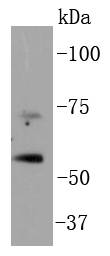
Fig1: Western blot analysis of p73 on Hela cell lysates using anti-p73 antibody at 1/1,000 dilution.Application
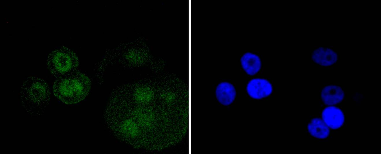
Fig2: ICC staining p73 in MCF-7 cells (green). The nuclear counter stain is DAPI (blue). Cells were fixed in paraformaldehyde, permeabilised with 0.25% Triton X100/PBS.Application
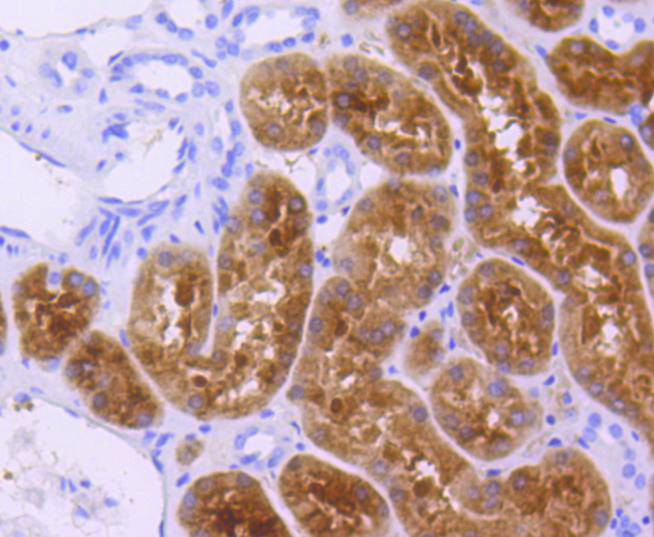
Fig3: Immunohistochemical analysis of paraffin-embedded human kidney tissue using anti-p73 antibody. Counter stained with hematoxylin.Application
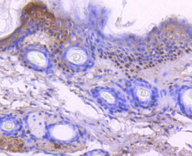
Fig4: Immunohistochemical analysis of paraffin-embedded mouse skin tissue using anti-p73 antibody. Counter stained with hematoxylin.Application
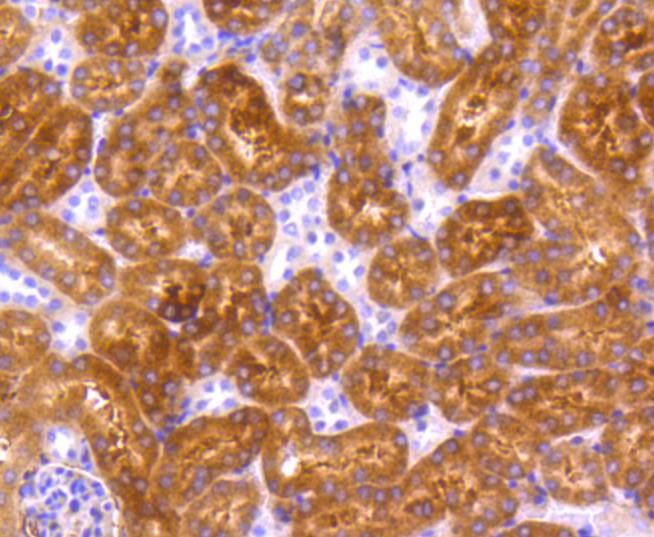
Fig5: Immunohistochemical analysis of paraffin-embedded mouse kidney tissue using anti-p73 antibody. Counter stained with hematoxylin.| Product Name | p73 [ST05-76] |
|---|---|
| Antibody Type | Primary Antibodies |
| Antigen Alias | p53 like transcription factor antibody p53 related protein antibody p53-like transcription factor antibody p53-related protein antibody p73 antibody P73_HUMAN antibody TP73 antibody Tumor protein p73 antibody |
| Product description | as. However, p73 does not appear to be activated by DNA damaging agents. The p73 isoform p73α inhibits drug-induced apoptosis in small cell lung carcinoma cells, while the p73 isoform p73β promotes it. p73α also prevents Bax activation, mitochondrial dysfunction, caspase activation and is able to reduce apoptosis induced by the BH3-only protein PUMA (p53 upregulated modulator of apoptosis). There is an equilibrium between p73α and p73β, demonstrated by the fact that p73α inhibits the pro-apoptotic effect of p73β. |
| Immunogen | recombinant protein |
| Clonality | Monoclonal |
|---|---|
| Isotype | IgG |
| Host Species | Recombinant rabbit |
| Tested Applications | WBICC/IFIHC |
| WB:1:1,000 ICC:1:50-1:200 IHC:1:50-1:200 |
|
| Species Reactivity | HumanMouse |
| Concentration | 1mg/ml |
| Alternative Names | p53 like transcription factor antibody p53 related protein antibody p53-like transcription factor antibody p53-related protein antibody p73 antibody P73_HUMAN antibody TP73 antibody Tumor protein p73 antibody |
|---|---|
| Molecular Weight(MW) | 70/60 kDa(12 isoforms) |
| Cellular Localization | Cytoplasm, Nucleus. |
Application
Fig1: Western blot analysis of p73 on Hela cell lysates using anti-p73 antibody at 1/1,000 dilution.
Application
Fig2: ICC staining p73 in MCF-7 cells (green). The nuclear counter stain is DAPI (blue). Cells were fixed in paraformaldehyde, permeabilised with 0.25% Triton X100/PBS.
Application
Fig3: Immunohistochemical analysis of paraffin-embedded human kidney tissue using anti-p73 antibody. Counter stained with hematoxylin.
Application
Fig4: Immunohistochemical analysis of paraffin-embedded mouse skin tissue using anti-p73 antibody. Counter stained with hematoxylin.
Application
Fig5: Immunohistochemical analysis of paraffin-embedded mouse kidney tissue using anti-p73 antibody. Counter stained with hematoxylin.| Positive Control | MCF-7, Hela, human kidney tissue, mouse skin tissue, mouse kidney tissue. |
|---|---|
| Application Notes | WB:1:1,000 ICC:1:50-1:200 IHC:1:50-1:200 |
| Form | Liquid |
|---|---|
| Storage Instructions | Store at +4℃ after thawing. Aliquot store at -20℃ or -80℃. Avoid repeated freeze / thaw cycles. |
| Storage Buffer | 1*TBS (pH7.4), 1%BSA, 40%Glycerol. Preservative: 0.05% Sodium Azide. |


