Application
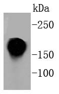
Fig1: Western blot analysis of CD11a on Jurkat cell lysates using anti-CD11a antibody at 1/1,000 dilution.Application
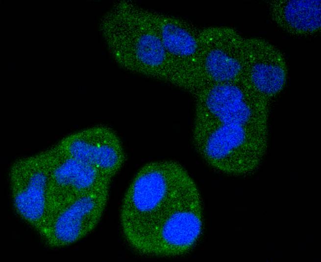
Fig2: ICC staining CD11a in Hela cells (green). The nuclear counter stain is DAPI (blue). Cells were fixed in paraformaldehyde, permeabilised with 0.25% Triton X100/PBS.Application
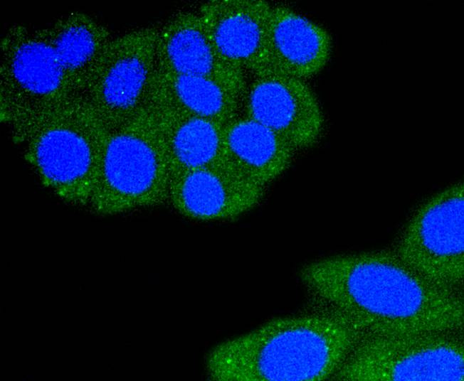
Fig3: ICC staining CD11a in HepG2 cells (green). The nuclear counter stain is DAPI (blue). Cells were fixed in paraformaldehyde, permeabilised with 0.25% Triton X100/PBS.Application
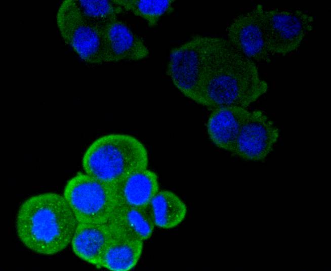
Fig4: ICC staining CD11a in SW480 cells (green). The nuclear counter stain is DAPI (blue). Cells were fixed in paraformaldehyde, permeabilised with 0.25% Triton X100/PBS.Application
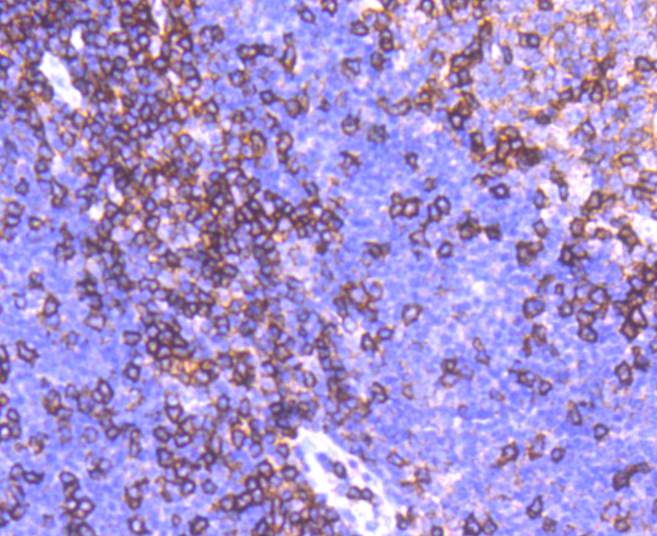
Fig5: Immunohistochemical analysis of paraffin-embedded human tonsil tissue using anti-CD11a antibody. Counter stained with hematoxylin.Application
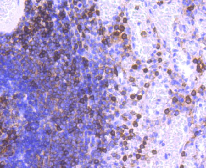
Fig6: Immunohistochemical analysis of paraffin-embedded human spleen tissue using anti-CD11a antibody. Counter stained with hematoxylin.Application
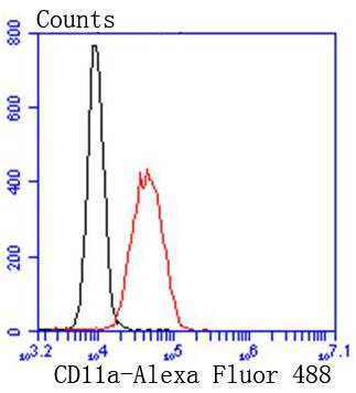
Fig7: Flow cytometric analysis of K562 cells with CD11a antibody at 1/50 dilution (red) compared with an unlabelled control (cells without incubation with primary antibody; black). Alexa Fluor 488-conjugated goat anti rabbit IgG was used as the secondary antibody.| Product Name | CD11a [SC56-01] |
|---|---|
| Antibody Type | Primary Antibodies |
| Antigen Alias | Lymphocyte function associated antigen, type 1, alpha subunit antibody Antigen CD11A (p180), lymphocyte function associated antigen 1, alpha polypeptide antibody Antigen CD11A antibody CD 11a antibody CD11 antigen-like family member A antibody CD11a antibody CD11a antigen antibody Integrin Alpha L antibody Integrin alpha-L antibody Integrin gene promoter antibody Integrin, alpha L (antigen CD11A (p180), lymphocyte function associated antigen 1; alpha polypeptide) antibody ITAL_HUMAN antibody Itgal antibody ITGAL protein antibody Leukocyte adhesion glycoprotein LFA 1 alpha chain antibody Leukocyte adhesion glycoprotein LFA-1 alpha chain antibody Leukocyte Adhesion Glycoprotein LFA1 Alpha Chain antibody Leukocyte function associated molecule 1 alpha chain antibody Leukocyte function-associated molecule 1 alpha chain antibody LFA 1 alpha (LFA1A) antibody LFA 1 alpha antibody LFA 1 antibody LFA 1A antibody LFA-1A antibody LFA1A antibody Ly15 antibody Ly21 antibody Lymphocyte function associated antigen 1 antibody Lymphocyte Function Associated Antigen Type 1 alpha antibody lymphocyte function-associated antigen 1, alpha polypeptide antibody p180 antibody |
| Product description | Integrins are heterodimers composed of noncovalently associated transmembrane a and b subunits. The 16 a and 8 b subunits heterodimerize to produce more than 20 different receptors. Most integrin receptors bind ligands that are components of the extracellular matrix, including Fibronectin, Collagen and Vitronectin. Certain integrins can also bind to soluble ligands such as Fibrinogen, or to counterreceptors on adjacent cells such as the intracellular adhesion molecules (ICAMs), leading to aggregation of cells. Ligands serve to cross-link or cluster integrins by binding to adjacent integrin receptors; both receptor clustering and ligand occupancy are necessary for the activation of integrin-mediated responses. In addition to mediating cell adhesion and cytoskeletal organization, integrins function as signaling receptors. Signals transduced by integrins play a role in many biological processes, including cell growth, differentiation, migration and apoptosis. |
| Immunogen | recombinant protein |
| Clonality | Monoclonal |
|---|---|
| Isotype | IgG |
| Host Species | Recombinant rabbit |
| Tested Applications | WBICC/IFIHCIPFC |
| WB:1:1,000-1:5,000 ICC:1:50-1:200 IHC:1:50-1:200 FC:1:50-1:100 |
|
| Species Reactivity | Human |
| Concentration | 1mg/ml |
| Alternative Names | Lymphocyte function associated antigen type 1 alpha subunit antibody Antigen CD11A (p180) lymphocyte function associated antigen 1 alpha polypeptide antibody Antigen CD11A antibody CD 11a antibody CD11 antigen-like family member A antibody CD11a antibody CD11a antigen antibody Integrin Alpha L antibody Integrin alpha-L antibody Integrin gene promoter antibody Integrin alpha L (antigen CD11A (p180) lymphocyte function associated antigen 1 alpha polypeptide) antibody ITAL_HUMAN antibody Itgal antibody ITGAL protein antibody Leukocyte adhesion glycoprotein LFA 1 alpha chain antibody Leukocyte adhesion glycoprotein LFA-1 alpha chain antibody Leukocyte Adhesion Glycoprotein LFA1 Alpha Chain antibody Leukocyte function associated molecule 1 alpha chain antibody Leukocyte function-associated molecule 1 alpha chain antibody LFA 1 alpha (LFA1A) antibody LFA 1 alpha antibody LFA 1 antibody LFA 1A antibody LFA-1A antibody LFA1A antibody Ly15 antibody Ly21 antibody Lymphocyte function associated antigen 1 antibody Lymphocyte Function Associated Antigen Type 1 alpha antibody lymphocyte function-associated antigen 1 alpha polypeptide antibody p180 antibody |
|---|---|
| Molecular Weight(MW) | 180 kDa |
| Cellular Localization | Membrane. |
| SwissProt ID | P20701 |
|---|
Application
Fig1: Western blot analysis of CD11a on Jurkat cell lysates using anti-CD11a antibody at 1/1,000 dilution.
Application
Fig2: ICC staining CD11a in Hela cells (green). The nuclear counter stain is DAPI (blue). Cells were fixed in paraformaldehyde, permeabilised with 0.25% Triton X100/PBS.
Application
Fig3: ICC staining CD11a in HepG2 cells (green). The nuclear counter stain is DAPI (blue). Cells were fixed in paraformaldehyde, permeabilised with 0.25% Triton X100/PBS.
Application
Fig4: ICC staining CD11a in SW480 cells (green). The nuclear counter stain is DAPI (blue). Cells were fixed in paraformaldehyde, permeabilised with 0.25% Triton X100/PBS.
Application
Fig5: Immunohistochemical analysis of paraffin-embedded human tonsil tissue using anti-CD11a antibody. Counter stained with hematoxylin.
Application
Fig6: Immunohistochemical analysis of paraffin-embedded human spleen tissue using anti-CD11a antibody. Counter stained with hematoxylin.
Application
Fig7: Flow cytometric analysis of K562 cells with CD11a antibody at 1/50 dilution (red) compared with an unlabelled control (cells without incubation with primary antibody; black). Alexa Fluor 488-conjugated goat anti rabbit IgG was used as the secondary antibody.| Positive Control | Jurkat, SW480, Hela, HepG2, human spleen tissue, human tonsil tissue. |
|---|---|
| Application Notes | WB:1:1,000-1:5,000 ICC:1:50-1:200 IHC:1:50-1:200 FC:1:50-1:100 |
| Form | Liquid |
|---|---|
| Storage Instructions | Store at +4℃ after thawing. Aliquot store at -20℃ or -80℃. Avoid repeated freeze / thaw cycles. |
| Storage Buffer | 1*TBS (pH7.4), 1%BSA, 40%Glycerol. Preservative: 0.05% Sodium Azide. |


