WB
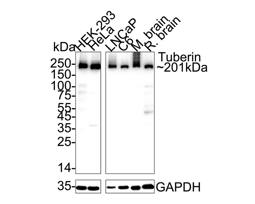
Western blot analysis of Tuberin on different lysates with Rabbit anti-Tuberin antibody at 1/1,000 dilution. Lane 1: HEK-293 cell lysate (20 µg/Lane), Lane 2: HeLa cell lysate (20 µg/Lane), Lane 3: LNCaP cell lysate (20 µg/Lane), Lane 4: C6 cell lysate (20 µg/Lane), Lane 5: Mouse brain tissue lysate (40 µg/Lane), Lane 6: Rat brain tissue lysate (40 µg/Lane), Exposure time: 25 seconds; 4-20% SDS-PAGE gel. Proteins were transferred to a PVDF membrane and blocked with 5% NFDM/TBST for 1 hour at room temperature. The primary antibody at 1/1,000 dilution was used in 5% NFDM/TBST at 4℃ overnight. Goat Anti-Rabbit IgG - HRP Secondary Antibody at 1/50,000 dilution was used for 1 hour at room temperature.IHC
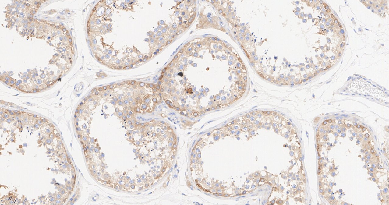
Immunohistochemical analysis of paraffin-embedded human testis tissue with Rabbit anti-Tuberin antibody at 1/200 dilution. The section was pre-treated using heat mediated antigen retrieval with Tris-EDTA buffer (pH 9.0) for 20 minutes. The tissues were blocked in 1% BSA for 20 minutes at room temperature, washed with ddH2O and PBS, and then probed with the primary antibody at 1/200 dilution for 1 hour at room temperature. The detection was performed using an HRP conjugated compact polymer system. DAB was used as the chromogen. Tissues were counterstained with hematoxylin and mounted with DPX.IHC
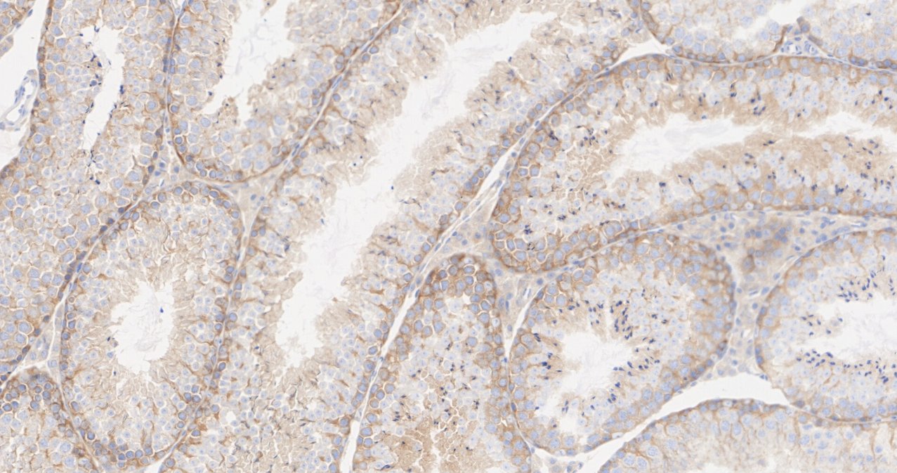
Immunohistochemical analysis of paraffin-embedded mouse testis tissue with Rabbit anti-Tuberin antibody at 1/200 dilution. The section was pre-treated using heat mediated antigen retrieval with Tris-EDTA buffer (pH 9.0) for 20 minutes. The tissues were blocked in 1% BSA for 20 minutes at room temperature, washed with ddH2O and PBS, and then probed with the primary antibody at 1/200 dilution for 1 hour at room temperature. The detection was performed using an HRP conjugated compact polymer system. DAB was used as the chromogen. Tissues were counterstained with hematoxylin and mounted with DPX.IHC
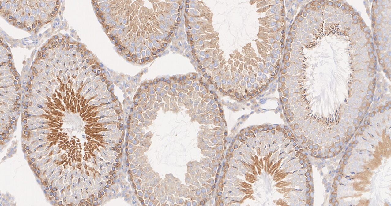
Immunohistochemical analysis of paraffin-embedded rat testis tissue with Rabbit anti-Tuberin antibody at 1/200 dilution. The section was pre-treated using heat mediated antigen retrieval with Tris-EDTA buffer (pH 9.0) for 20 minutes. The tissues were blocked in 1% BSA for 20 minutes at room temperature, washed with ddH2O and PBS, and then probed with the primary antibody at 1/200 dilution for 1 hour at room temperature. The detection was performed using an HRP conjugated compact polymer system. DAB was used as the chromogen. Tissues were counterstained with hematoxylin and mounted with DPX.FC
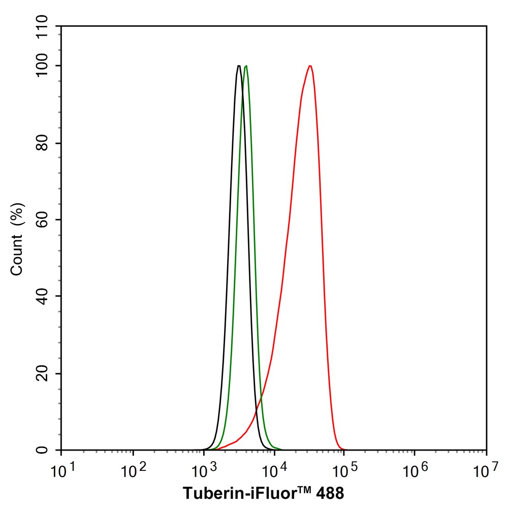
Flow cytometric analysis of HeLa cells labeling Tuberin. Cells were fixed and permeabilized. Then stained with the primary antibody (1:1000) (red) compared with Rabbit IgG Isotype Control (green). After incubation of the primary antibody at +4℃ for an hour, the cells were stained with a iFluor™ 488 conjugate-Goat anti-Rabbit IgG Secondary antibody at 1/1,000 dilution for 30 minutes at +4℃. Unlabelled sample was used as a control (cells without incubation with primary antibody; black).| Product Name | Tuberin Recombinant Rabbit Monoclonal Antibody |
|---|---|
| Antibody Type | Primary Antibodies |
| Product description | Tuberous sclerosis complex (TSC) is an autosomal dominant genetic disorder characterized by mental retardation and the widespread development of distinctive tumors termed hamartomas. Two different genetic loci have been linked to TSC; one of these loci, the tuberous sclerosis-2 gene (TSC2), encodes a protein called tuberin and the other loci, tuberous sclerosis-1 gene (TSC1), encodes a protein called hamartin. Tuberin and hamartin interact with each other forming a cystoplasmic complex. Hamartin interacts with the ezrin-radixin-moesin (ERM) family of actin-binding proteins and inhibition of hamartin activity results in loss of cell adhesion. Hamartin is present in most adult tissues with strong expression in brain, heart, and kidney. |
| Immunogen | Synthetic peptide within Human Tuberin aa 1760-1807 / 1807. |
| Clonality | Monoclonal |
|---|---|
| Isotype | IgG |
| Host Species | Recombinant rabbit |
| Tested Applications | FCIHCWB |
| WB:1:500-1:2000 IHC:1:200 FC:1:1000 |
|
| Species Reactivity | HumanMouseRat |
| Concentration | 1mg/ml |
| Purification | Protein A |
| Alternative Names | FLJ43106 antibody
LAM antibody
OTTHUMP00000158940 antibody
OTTHUMP00000198394 antibody
OTTHUMP00000198395 antibody
PPP1R160 antibody
Protein phosphatase 1 regulatory subunit 160 antibody tsc2 antibody TSC2_HUMAN antibody TSC4 antibody TSC4 gene formerly antibody TSC4 formerly antibody Tuberin antibody Tuberous sclerosis 2 antibody Tuberous sclerosis 2 protein antibody Tuberous sclerosis 2 protein homolog antibody |
|---|---|
| Molecular Weight(MW) | 201 kDa |
| Cellular Localization | Cytoplasm, Membrane. |
WB
Western blot analysis of Tuberin on different lysates with Rabbit anti-Tuberin antibody at 1/1,000 dilution. Lane 1: HEK-293 cell lysate (20 µg/Lane), Lane 2: HeLa cell lysate (20 µg/Lane), Lane 3: LNCaP cell lysate (20 µg/Lane), Lane 4: C6 cell lysate (20 µg/Lane), Lane 5: Mouse brain tissue lysate (40 µg/Lane), Lane 6: Rat brain tissue lysate (40 µg/Lane), Exposure time: 25 seconds; 4-20% SDS-PAGE gel. Proteins were transferred to a PVDF membrane and blocked with 5% NFDM/TBST for 1 hour at room temperature. The primary antibody at 1/1,000 dilution was used in 5% NFDM/TBST at 4℃ overnight. Goat Anti-Rabbit IgG - HRP Secondary Antibody at 1/50,000 dilution was used for 1 hour at room temperature.
IHC
Immunohistochemical analysis of paraffin-embedded human testis tissue with Rabbit anti-Tuberin antibody at 1/200 dilution. The section was pre-treated using heat mediated antigen retrieval with Tris-EDTA buffer (pH 9.0) for 20 minutes. The tissues were blocked in 1% BSA for 20 minutes at room temperature, washed with ddH2O and PBS, and then probed with the primary antibody at 1/200 dilution for 1 hour at room temperature. The detection was performed using an HRP conjugated compact polymer system. DAB was used as the chromogen. Tissues were counterstained with hematoxylin and mounted with DPX.
IHC
Immunohistochemical analysis of paraffin-embedded mouse testis tissue with Rabbit anti-Tuberin antibody at 1/200 dilution. The section was pre-treated using heat mediated antigen retrieval with Tris-EDTA buffer (pH 9.0) for 20 minutes. The tissues were blocked in 1% BSA for 20 minutes at room temperature, washed with ddH2O and PBS, and then probed with the primary antibody at 1/200 dilution for 1 hour at room temperature. The detection was performed using an HRP conjugated compact polymer system. DAB was used as the chromogen. Tissues were counterstained with hematoxylin and mounted with DPX.
IHC
Immunohistochemical analysis of paraffin-embedded rat testis tissue with Rabbit anti-Tuberin antibody at 1/200 dilution. The section was pre-treated using heat mediated antigen retrieval with Tris-EDTA buffer (pH 9.0) for 20 minutes. The tissues were blocked in 1% BSA for 20 minutes at room temperature, washed with ddH2O and PBS, and then probed with the primary antibody at 1/200 dilution for 1 hour at room temperature. The detection was performed using an HRP conjugated compact polymer system. DAB was used as the chromogen. Tissues were counterstained with hematoxylin and mounted with DPX.
FC
Flow cytometric analysis of HeLa cells labeling Tuberin. Cells were fixed and permeabilized. Then stained with the primary antibody (1:1000) (red) compared with Rabbit IgG Isotype Control (green). After incubation of the primary antibody at +4℃ for an hour, the cells were stained with a iFluor™ 488 conjugate-Goat anti-Rabbit IgG Secondary antibody at 1/1,000 dilution for 30 minutes at +4℃. Unlabelled sample was used as a control (cells without incubation with primary antibody; black).| Application Notes | WB:1:500-1:2000 IHC:1:200 FC:1:1000 |
|---|
| Form | Liquid |
|---|---|
| Storage Instructions | Store at +4℃ after thawing. Aliquot store at -20℃ or -80℃. Avoid repeated freeze / thaw cycles. |
| Storage Buffer | 1*TBS (pH7.4), 1%BSA, 40%Glycerol. Preservative: 0.05% Sodium Azide. |


