WB
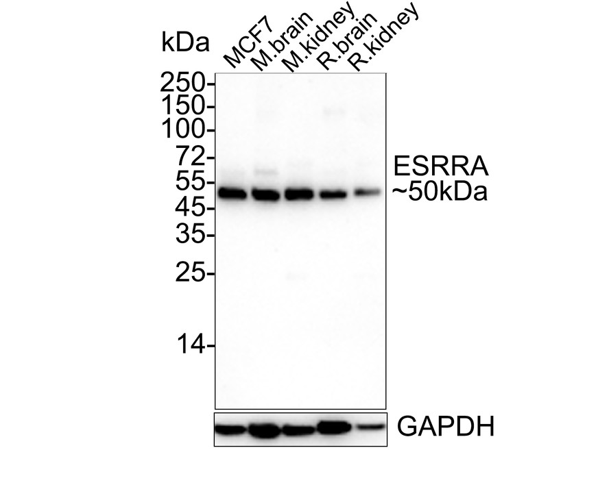
Western blot analysis of Estrogen Related Receptor alpha on different lysates with Rabbit anti-Estrogen Related Receptor alpha antibody at 1/2,000 dilution. Lane 1: MCF-7 cell lysate, Lane 2: mouse brain tissue lysate, Lane 3: mouse kidney tissue lysate, Lane 4: rat brain tissue lysate, Lane 5: rat kidney tissue lysate, Lysates/proteins at 30 µg/Lane. Exposure time: 43 seconds; 4-20% SDS-PAGE gel. Proteins were transferred to a PVDF membrane and blocked with 5% NFDM/TBST for 1 hour at room temperature. The primary antibody at 1/1,000 dilution was used in 5% NFDM/TBST at 4℃ overnight. Goat Anti-Rabbit IgG - HRP Secondary Antibody at 1/50,000 dilution was used for 1 hour at room temperature.FC
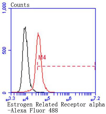
Flow cytometric analysis of Hela cells with Estrogen Related Receptor alpha antibody at 1/50 dilution (red) compared with an unlabelled control (cells without incubation with primary antibody; black). Alexa Fluor 488-conjugated goat anti rabbit IgG was used as the secondary antibody.IHC
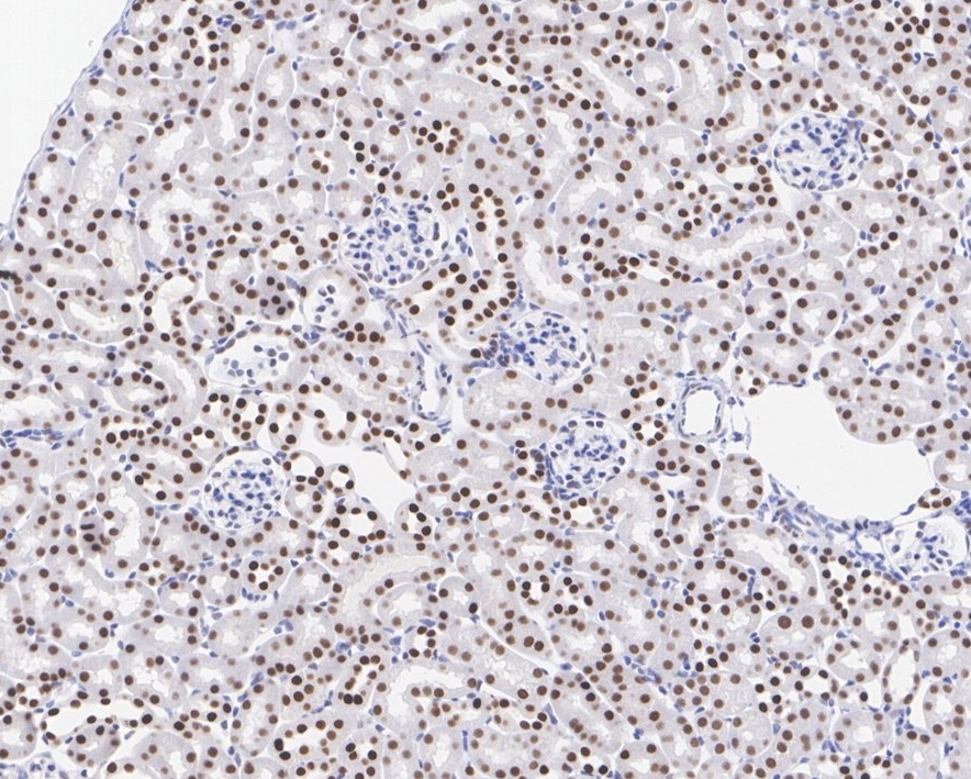
Immunohistochemical analysis of paraffin-embedded mouse kidney tissue with Rabbit anti-Estrogen Related Receptor alpha antibody at 1/1,000 dilution. The section was pre-treated using heat mediated antigen retrieval with sodium citrate buffer (pH 6.0) for 2 minutes. The tissues were blocked in 1% BSA for 20 minutes at room temperature, washed with ddH2O and PBS, and then probed with the primary antibody at 1/1,000 dilution for 1 hour at room temperature. The detection was performed using an HRP conjugated compact polymer system. DAB was used as the chromogen. Tissues were counterstained with hematoxylin and mounted with DPX.| Product Name | Estrogen Related Receptor alpha Recombinant Rabbit Monoclonal Antibody |
|---|---|
| Antibody Type | Primary Antibodies |
| Product description | Estrogen related receptor alpha (ERRα) is a nuclear receptor in the superfamily of ligand-regulated transcription factors and is a member of the NR3B orphan nuclear receptor subgroup (consisting of α, β and γ). ERRα plays a role in modulating the estrogen signaling pathway. In addition, the expression of ERRα has been shown to increase during fasting and cold exposure. ERRα may be important for regulating mitochondrial biogenesis and oxidative metabolism by acting directly on genes necessary for mitochondrial function. Mice lacking ERRα are unable to maintain their body temperature in the cold. ERRα may also be involved in the maintenance and formation of cartilage. This information could be useful in finding therapeutic agents for a variety of diseases affecting the joints. |
| Immunogen | Synthetic peptide within Human Estrogen Related Receptor alpha aa 1-49 / 423. |
| Clonality | monoclonal |
|---|---|
| Isotype | IgG |
| Host Species | Recombinant rabbit |
| Tested Applications | FCIHCWB |
| WB:1:1000-1:2000 FC:1:50-1:100 IHC:1:1000 |
|
| Species Reactivity | HumanMouseRat |
| Concentration | 1mg/ |
| Purification | Protein A |
| Alternative Names | Err 1 antibody ERR a antibody ERR alpha antibody ERR-alpha antibody Err1 antibody ERR1 protein antibody ERR1_HUMAN antibody ERRa antibody ERRalpha antibody ESRL 1 antibody ESRL1 antibody ESRR A antibody Esrra antibody Estrogen receptor like 1 antibody Estrogen receptor related 1 antibody estrogen receptor related receptor alpha antibody Estrogen receptor-like 1 antibody Estrogen related receptor alpha antibody Estrogen-related receptor alpha antibody Estrra antibody hERR1 antibody NR3B1 antibody Nuclear receptor subfamily 3 group B member 1 antibody Steroid hormone receptor ERR1 antibody |
|---|---|
| Molecular Weight(MW) | 46kDa(Observed band size: 50kDa) |
| Cellular Localization | Nucleus. |
WB
Western blot analysis of Estrogen Related Receptor alpha on different lysates with Rabbit anti-Estrogen Related Receptor alpha antibody at 1/2,000 dilution. Lane 1: MCF-7 cell lysate, Lane 2: mouse brain tissue lysate, Lane 3: mouse kidney tissue lysate, Lane 4: rat brain tissue lysate, Lane 5: rat kidney tissue lysate, Lysates/proteins at 30 µg/Lane. Exposure time: 43 seconds; 4-20% SDS-PAGE gel. Proteins were transferred to a PVDF membrane and blocked with 5% NFDM/TBST for 1 hour at room temperature. The primary antibody at 1/1,000 dilution was used in 5% NFDM/TBST at 4℃ overnight. Goat Anti-Rabbit IgG - HRP Secondary Antibody at 1/50,000 dilution was used for 1 hour at room temperature.
FC
Flow cytometric analysis of Hela cells with Estrogen Related Receptor alpha antibody at 1/50 dilution (red) compared with an unlabelled control (cells without incubation with primary antibody; black). Alexa Fluor 488-conjugated goat anti rabbit IgG was used as the secondary antibody.
IHC
Immunohistochemical analysis of paraffin-embedded mouse kidney tissue with Rabbit anti-Estrogen Related Receptor alpha antibody at 1/1,000 dilution. The section was pre-treated using heat mediated antigen retrieval with sodium citrate buffer (pH 6.0) for 2 minutes. The tissues were blocked in 1% BSA for 20 minutes at room temperature, washed with ddH2O and PBS, and then probed with the primary antibody at 1/1,000 dilution for 1 hour at room temperature. The detection was performed using an HRP conjugated compact polymer system. DAB was used as the chromogen. Tissues were counterstained with hematoxylin and mounted with DPX.| Positive Control | MCF-7. |
|---|---|
| Application Notes | WB:1:1000-1:2000 FC:1:50-1:100 IHC:1:1000 |
| Form | Liquid |
|---|---|
| Storage Instructions | Store at +4℃ after thawing. Aliquot store at -20℃ or -80℃. Avoid repeated freeze / thaw cycles. |
| Storage Buffer | 1*TBS (pH7.4), 1%BSA, 40%Glycerol. Preservative: 0.05% Sodium Azide. |


