WB
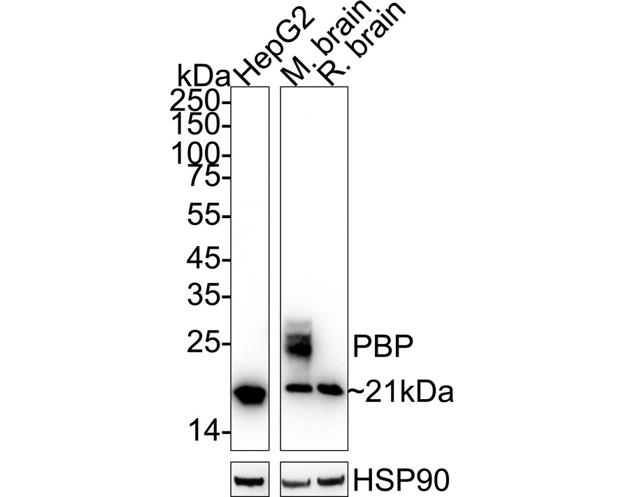
Western blot analysis of PBP on different lysates with Rabbit anti-PBP antibody at 1/1,000 dilution. Lane 1: HepG2 cell lysate (20 µg/Lane), Lane 2: Mouse brain tissue lysate (40 µg/Lane), Lane 3: Rat brain tissue lysate (40 µg/Lane), Exposure time: 59 seconds; 4-20% SDS-PAGE gel. Proteins were transferred to a PVDF membrane and blocked with 5% NFDM/TBST for 1 hour at room temperature. The primary antibody at 1/1,000 dilution was used in 5% NFDM/TBST at 4℃ overnight. Goat Anti-Rabbit IgG - HRP Secondary Antibody at 1/50,000 dilution was used for 1 hour at room temperature.ICC/IF
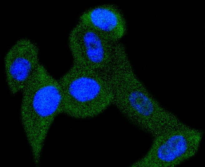
ICC staining PBP in PC-3M cells (green). The nuclear counter stain is DAPI (blue). Cells were fixed in paraformaldehyde, permeabilised with 0.25% Triton X100/PBS.IHC
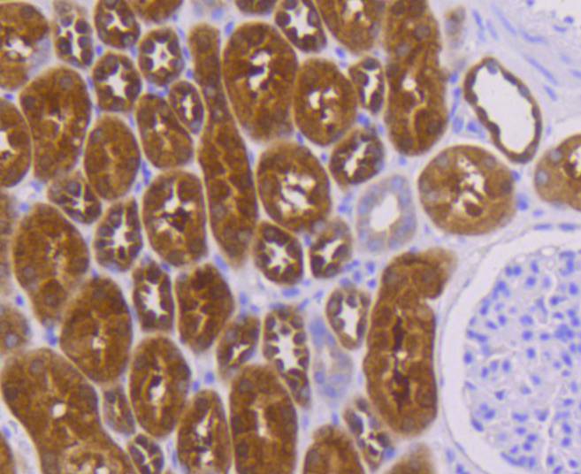
Immunohistochemical analysis of paraffin-embedded human kidney tissue using anti-PBP antibody. Counter stained with hematoxylin.IHC
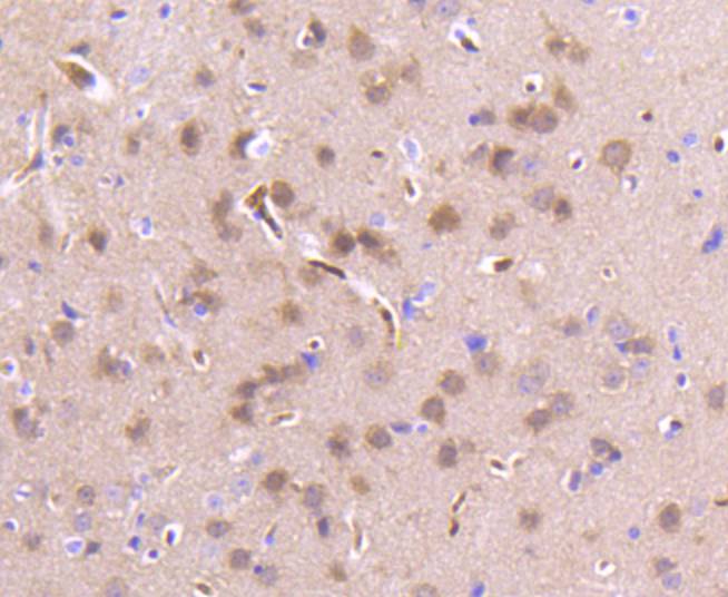
Immunohistochemical analysis of paraffin-embedded mouse brain tissue using anti-PBP antibody. Counter stained with hematoxylin.IHC
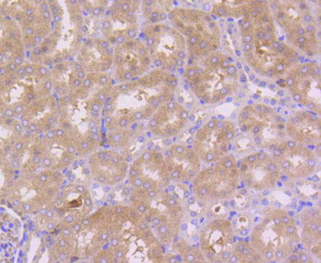
Immunohistochemical analysis of paraffin-embedded mouse kidney tissue using anti-PBP antibody. Counter stained with hematoxylin.| Product Name | PBP Recombinant Rabbit Monoclonal Antibody |
|---|---|
| Antibody Type | Primary Antibodies |
| Product description | Members of the a-chemokine subfamily of inducible, secreted, pro-inflammatory cytokines contain a similar motif, in which the first two cysteine residues are separated by a single residue (Cys-X-Cys), and are also chemotactic for neutrophils. The platelet basic protein (PBP), a member of the a(lpha)-chemokine family, resides in the a(lpha)-granules of platelets and is released upon their activation. Proteolytic cleavage of the amino terminus of PBP leads to the generation of several peptides, which include mature PBP, connective tissue-activating peptide III (CTAP III, also designated low affinity platelet factor IV (LA-PF4)), b-thromboglobulin (b-TG), and neutrophil-activating peptide 2 (NAP-2). PBP and its N-truncated derivatives mediate inflammation and wound healing. Specifically, NAP-2 activates chemotaxis and degranulation in neutrophils during inflammation. The gene encoding human PBP maps to chromosome 4q12-q13. |
| Immunogen | Synthetic peptide within Human PBP aa 53-96 / 187. |
| Clonality | Monoclonal |
|---|---|
| Isotype | IgG |
| Host Species | Recombinant rabbit |
| Tested Applications | ICC/IFIHCWB |
| WB:1:1000-1:5000 IHC:1:50-1:200 ICC/IF:1:50-1:200 |
|
| Species Reactivity | HumanMouseRatZebrafish |
| Concentration | 1mg/ml |
| Purification | Protein A |
| Alternative Names | Epididymis luminal protein 210 antibody Epididymis secretory protein Li 34 antibody Epididymis secretory protein Li 96 antibody HCNP antibody HCNPpp antibody HEL 210 antibody HEL S 34 antibody HEL S 96 antibody Hippocampal cholinergic neurostimulating peptide antibody Neuropolypeptide h3 antibody PBP antibody PEBP antibody PEBP 1 antibody PEBP-1 antibody Pebp1 antibody PEBP1_HUMAN antibody Phosphatidylethanolamine binding protein antibody Phosphatidylethanolamine binding protein 1 antibody Prostatic binding protein antibody Prostatic-binding protein antibody R kip antibody Raf kinase inhibitor protein antibody Raf kinase inhibitory protein antibody RKIP antibody |
|---|---|
| Molecular Weight(MW) | 21 kDa |
| Cellular Localization | Cytoplasm. |
WB
Western blot analysis of PBP on different lysates with Rabbit anti-PBP antibody at 1/1,000 dilution. Lane 1: HepG2 cell lysate (20 µg/Lane), Lane 2: Mouse brain tissue lysate (40 µg/Lane), Lane 3: Rat brain tissue lysate (40 µg/Lane), Exposure time: 59 seconds; 4-20% SDS-PAGE gel. Proteins were transferred to a PVDF membrane and blocked with 5% NFDM/TBST for 1 hour at room temperature. The primary antibody at 1/1,000 dilution was used in 5% NFDM/TBST at 4℃ overnight. Goat Anti-Rabbit IgG - HRP Secondary Antibody at 1/50,000 dilution was used for 1 hour at room temperature.
ICC/IF
ICC staining PBP in PC-3M cells (green). The nuclear counter stain is DAPI (blue). Cells were fixed in paraformaldehyde, permeabilised with 0.25% Triton X100/PBS.
IHC
Immunohistochemical analysis of paraffin-embedded human kidney tissue using anti-PBP antibody. Counter stained with hematoxylin.
IHC
Immunohistochemical analysis of paraffin-embedded mouse brain tissue using anti-PBP antibody. Counter stained with hematoxylin.
IHC
Immunohistochemical analysis of paraffin-embedded mouse kidney tissue using anti-PBP antibody. Counter stained with hematoxylin.| Application Notes | WB:1:1000-1:5000 IHC:1:50-1:200 ICC/IF:1:50-1:200 |
|---|
| Form | Liquid |
|---|---|
| Storage Instructions | Store at +4℃ after thawing. Aliquot store at -20℃ or -80℃. Avoid repeated freeze / thaw cycles. |
| Storage Buffer | 1*TBS (pH7.4), 1%BSA, 40%Glycerol. Preservative: 0.05% Sodium Azide. |


