WB
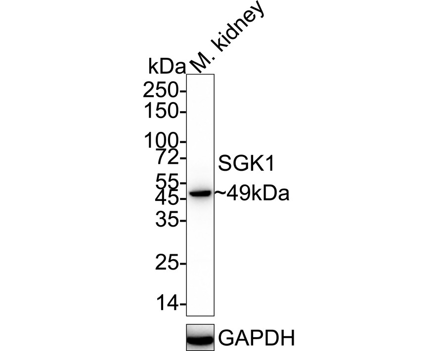
Western blot analysis of SGK1 on mouse kidney tissue lysates with Rabbit anti-SGK1 antibody at 1/500 dilution. Lysates/proteins at 40 µg/Lane. Exposure time: 30 seconds; 4-20% SDS-PAGE gel. Proteins were transferred to a PVDF membrane and blocked with 5% NFDM/TBST for 1 hour at room temperature. The primary antibody at 1/500 dilution was used in 5% NFDM/TBST at 4℃ overnight. Goat Anti-Rabbit IgG - HRP Secondary Antibody at 1/50,000 dilution was used for 1 hour at room temperature.IHC
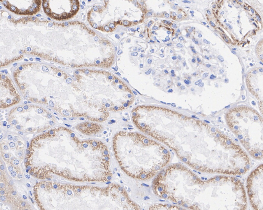
Immunohistochemical analysis of paraffin-embedded human kidney tissue with Rabbit anti-SGK1 antibody at 1/1,000 dilution. The section was pre-treated using heat mediated antigen retrieval with Tris-EDTA buffer (pH 9.0) for 20 minutes. The tissues were blocked in 1% BSA for 20 minutes at room temperature, washed with ddH2O and PBS, and then probed with the primary antibody at 1/1,000 dilution for 1 hour at room temperature. The detection was performed using an HRP conjugated compact polymer system. DAB was used as the chromogen. Tissues were counterstained with hematoxylin and mounted with DPX.IHC
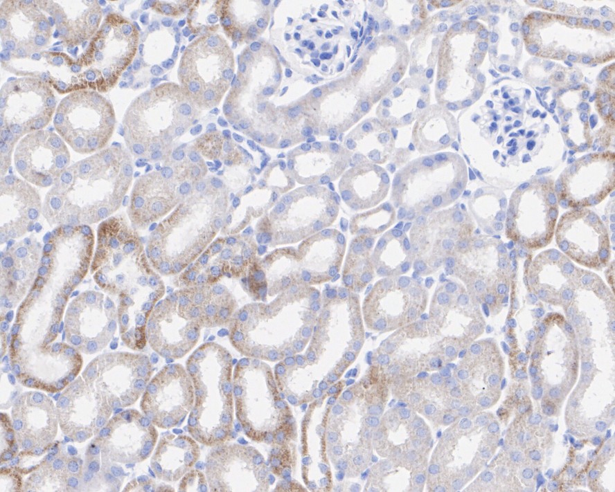
Immunohistochemical analysis of paraffin-embedded mouse kidney tissue with Rabbit anti-SGK1 antibody at 1/1,000 dilution. The section was pre-treated using heat mediated antigen retrieval with Tris-EDTA buffer (pH 9.0) for 20 minutes. The tissues were blocked in 1% BSA for 20 minutes at room temperature, washed with ddH2O and PBS, and then probed with the primary antibody at 1/1,000 dilution for 1 hour at room temperature. The detection was performed using an HRP conjugated compact polymer system. DAB was used as the chromogen. Tissues were counterstained with hematoxylin and mounted with DPX.IHC
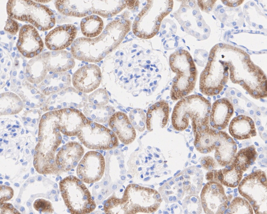
Immunohistochemical analysis of paraffin-embedded rat kidney tissue with Rabbit anti-SGK1 antibody at 1/1,000 dilution. The section was pre-treated using heat mediated antigen retrieval with Tris-EDTA buffer (pH 9.0) for 20 minutes. The tissues were blocked in 1% BSA for 20 minutes at room temperature, washed with ddH2O and PBS, and then probed with the primary antibody at 1/1,000 dilution for 1 hour at room temperature. The detection was performed using an HRP conjugated compact polymer system. DAB was used as the chromogen. Tissues were counterstained with hematoxylin and mounted with DPX.FC
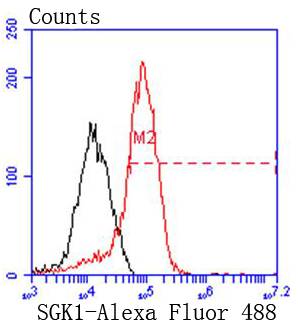
Flow cytometric analysis of 293 cells with SGK1 antibody at 1/50 dilution (red) compared with an unlabelled control (cells without incubation with primary antibody; black). Alexa Fluor 488-conjugated goat anti rabbit IgG was used as the secondary antibody.| Product Name | SGK1 Recombinant Rabbit Monoclonal Antibody |
|---|---|
| Antibody Type | Primary Antibodies |
| Product description | Serum- and glucocorticoid-regulated kinase (SGK), also known as SGK1, is a serine/threonine protein kinase and a member of the "AGC" subfamily, which includes protein kinases A, G, and C. SGK plays an important role in activating certain potassium, sodium, and chloride channels, suggesting an involvement in the regulation of processes such as cell survival, neuronal excitability, and renal sodium excretion. SGK contains a catalytic domain, which is most similar to Akt1 (also known as protein kinase B or PKB). SGK is a downstream target of PI 3-kinase-stimulated growth factor signaling, with 3-phosphoinositide-dependent protein kinase 1 (PDK1) capable of phosphorylating the activation-loop of SGK at Threonine-256. The adrenal corticosteroid hormone, Aldosterone, induces the transcription of SGK, which mediates Na + transport by stimulating epithelial sodium channel activity. The SGK promoter contains a glucocorticoid response element and an SP-1 regulatory element, and is a transcriptional target for p53. SGK is also a component of the p38 MAPK-mediated response to hyperosmotic stress. The human SGK gene maps to chromosome 6q23 and encodes the 431-amino acid SGK protein. |
| Immunogen | Synthetic peptide within human SGK1 aa 40-80. |
| Clonality | Monoclonal |
|---|---|
| Isotype | IgG |
| Host Species | Recombinant rabbit |
| Tested Applications | FCIHCWB |
| WB:1:500-1:1000 IHC:1:50-1:200 FC:1:50-1:100 |
|
| Species Reactivity | HumanMouseRat |
| Concentration | 1mg/ml |
| Purification | Protein A |
| Alternative Names | OTTHUMP00000017247 antibody Serine/threonine protein kinase SGK antibody Serine/threonine protein kinase Sgk1 antibody Serine/threonine-protein kinase Sgk1 antibody Serum and glucocorticoid regulated kinase antibody Serum/glucocorticoid regulated kinase 1 antibody Serum/glucocorticoid regulated kinase antibody Serum/glucocorticoid-regulated kinase 1 antibody SGK 1 antibody SGK antibody SGK1 antibody Sgk1 variant i3 antibody SGK1_HUMAN antibody |
|---|---|
| Molecular Weight(MW) | 49 kDa |
| Cellular Localization | Cytoplasm, Nucleus, Cell membrane, Mitochondrion. |
WB
Western blot analysis of SGK1 on mouse kidney tissue lysates with Rabbit anti-SGK1 antibody at 1/500 dilution. Lysates/proteins at 40 µg/Lane. Exposure time: 30 seconds; 4-20% SDS-PAGE gel. Proteins were transferred to a PVDF membrane and blocked with 5% NFDM/TBST for 1 hour at room temperature. The primary antibody at 1/500 dilution was used in 5% NFDM/TBST at 4℃ overnight. Goat Anti-Rabbit IgG - HRP Secondary Antibody at 1/50,000 dilution was used for 1 hour at room temperature.
IHC
Immunohistochemical analysis of paraffin-embedded human kidney tissue with Rabbit anti-SGK1 antibody at 1/1,000 dilution. The section was pre-treated using heat mediated antigen retrieval with Tris-EDTA buffer (pH 9.0) for 20 minutes. The tissues were blocked in 1% BSA for 20 minutes at room temperature, washed with ddH2O and PBS, and then probed with the primary antibody at 1/1,000 dilution for 1 hour at room temperature. The detection was performed using an HRP conjugated compact polymer system. DAB was used as the chromogen. Tissues were counterstained with hematoxylin and mounted with DPX.
IHC
Immunohistochemical analysis of paraffin-embedded mouse kidney tissue with Rabbit anti-SGK1 antibody at 1/1,000 dilution. The section was pre-treated using heat mediated antigen retrieval with Tris-EDTA buffer (pH 9.0) for 20 minutes. The tissues were blocked in 1% BSA for 20 minutes at room temperature, washed with ddH2O and PBS, and then probed with the primary antibody at 1/1,000 dilution for 1 hour at room temperature. The detection was performed using an HRP conjugated compact polymer system. DAB was used as the chromogen. Tissues were counterstained with hematoxylin and mounted with DPX.
IHC
Immunohistochemical analysis of paraffin-embedded rat kidney tissue with Rabbit anti-SGK1 antibody at 1/1,000 dilution. The section was pre-treated using heat mediated antigen retrieval with Tris-EDTA buffer (pH 9.0) for 20 minutes. The tissues were blocked in 1% BSA for 20 minutes at room temperature, washed with ddH2O and PBS, and then probed with the primary antibody at 1/1,000 dilution for 1 hour at room temperature. The detection was performed using an HRP conjugated compact polymer system. DAB was used as the chromogen. Tissues were counterstained with hematoxylin and mounted with DPX.
FC
Flow cytometric analysis of 293 cells with SGK1 antibody at 1/50 dilution (red) compared with an unlabelled control (cells without incubation with primary antibody; black). Alexa Fluor 488-conjugated goat anti rabbit IgG was used as the secondary antibody.| Application Notes | WB:1:500-1:1000 IHC:1:50-1:200 FC:1:50-1:100 |
|---|
| Form | Liquid |
|---|---|
| Storage Instructions | Store at +4℃ after thawing. Aliquot store at -20℃ or -80℃. Avoid repeated freeze / thaw cycles. |
| Storage Buffer | 1*TBS (pH7.4), 1%BSA, 40%Glycerol. Preservative: 0.05% Sodium Azide. |


