Application
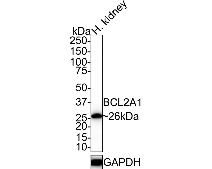
Fig1: Western blot analysis of BCL2A1 on human kidney tissue lysates with Rabbit anti-BCL2A1 antibody at 1/500 dilution. Lysates/proteins at 40 µg/Lane. 4-20% SDS-PAGE gel. Proteins were transferred to a PVDF membrane and blocked with 5% NFDM/TBST for 1 hour at room temperature. The primary antibody at 1/500 dilution was used in 5% NFDM/TBST at room temperature for 2 hours. Goat Anti-Rabbit IgG - HRP Secondary Antibody at 1:100,000 dilution was used for 1 hour at room temperature.Application
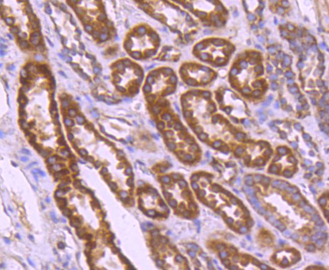
Fig2: Immunohistochemical analysis of paraffin-embedded human kidney tissue using anti- BCL2A1 antibody. Counter stained with hematoxylin.Application
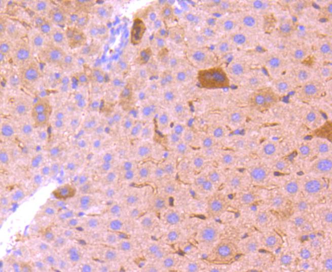
Fig3: Immunohistochemical analysis of paraffin-embedded mouse liver tissue using anti- BCL2A1 antibody. Counter stained with hematoxylin.Application
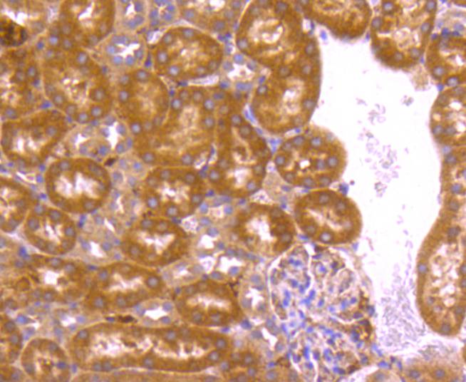
Fig4: Immunohistochemical analysis of paraffin-embedded mouse kidney tissue using anti- BCL2A1 antibody. Counter stained with hematoxylin.Application
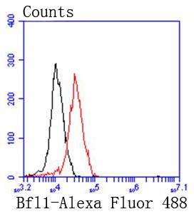
Fig5: Flow cytometric analysis of 293 cells with Bfl1(BCL2A1) antibody at 1/50 dilution (red) compared with an unlabelled control (cells without incubation with primary antibody; black). Alexa Fluor 488-conjugated goat anti rabbit IgG was used as the secondary antibody.| Product Name | BCL2A1 Recombinant Rabbit Monoclonal Antibody |
|---|---|
| Antibody Type | Primary Antibodies |
| Product description | The Bcl-2 family of proteins is characterized by its ability to modulate cell death under a broad range of physiological conditions. Bcl-2 and Bcl-xL function to inhibit apoptosis while other members of the Bcl-2 family, Bax, Bad, Bak and Bcl-xS, oppose death-suppressing effects. An additional member of the family, A1 (also designated Bfl-1), dimerizes with both Bcl-2 and Bax and has been identified as a hematopoietic- specific, early inducible gene. While A1 demonstrates life promoting properties similar to those of Bcl-2, its function may be more temporally regulated during myeloid differentiation and dependent on additional growth stimuli to confer its life promoting properties. A1 is abundantly expressed in bone marrow and at low levels in other tissues. There is evidence that a correlation exists between a high expression of the A1 gene product and stomach cancer. |
| Immunogen | Synthetic peptide within Human BCL2A1 aa 2-48 / 175. |
| Clonality | monoclonal |
|---|---|
| Isotype | IgG |
| Host Species | Recombinant rabbit |
| Tested Applications | FCIHCWB |
| WB:1:1,000 IHC:1:50-1:200: FC:1:50-1:100 |
|
| Species Reactivity | HumanMouseRat |
| Concentration | 1mg/ml |
| Purification | Protein A |
| Alternative Names | ACC 1 antibody ACC 2 antibody B2LA1_HUMAN antibody Bcl 2 related protein A1 antibody Bcl-2-like protein 5 antibody Bcl-2-related protein A1 antibody BCL2 related protein A1 antibody Bcl2-L-5 antibody BCL2A1 antibody BCL2L5 antibody BFL1 antibody GRS antibody HBPA1 antibody Hematopoietic BCL2 related protein A1 antibody Hemopoietic specific early response protein antibody Hemopoietic-specific early response protein antibody Protein BFL 1 antibody Protein BFL-1 antibody Protein GRS antibody |
|---|---|
| Molecular Weight(MW) | 20kDa(Observed band size: 26kDa) |
| Cellular Localization | Cytoplasm. |
Application
Fig1: Western blot analysis of BCL2A1 on human kidney tissue lysates with Rabbit anti-BCL2A1 antibody at 1/500 dilution. Lysates/proteins at 40 µg/Lane. 4-20% SDS-PAGE gel. Proteins were transferred to a PVDF membrane and blocked with 5% NFDM/TBST for 1 hour at room temperature. The primary antibody at 1/500 dilution was used in 5% NFDM/TBST at room temperature for 2 hours. Goat Anti-Rabbit IgG - HRP Secondary Antibody at 1:100,000 dilution was used for 1 hour at room temperature.
Application
Fig2: Immunohistochemical analysis of paraffin-embedded human kidney tissue using anti- BCL2A1 antibody. Counter stained with hematoxylin.
Application
Fig3: Immunohistochemical analysis of paraffin-embedded mouse liver tissue using anti- BCL2A1 antibody. Counter stained with hematoxylin.
Application
Fig4: Immunohistochemical analysis of paraffin-embedded mouse kidney tissue using anti- BCL2A1 antibody. Counter stained with hematoxylin.
Application
Fig5: Flow cytometric analysis of 293 cells with Bfl1(BCL2A1) antibody at 1/50 dilution (red) compared with an unlabelled control (cells without incubation with primary antibody; black). Alexa Fluor 488-conjugated goat anti rabbit IgG was used as the secondary antibody.| Positive Control | Human tonsil tissue, human kidney tissue, mouse liver tissue, mouse kidney tissue. |
|---|---|
| Application Notes | WB:1:1,000 IHC:1:50-1:200: FC:1:50-1:100 |
| Form | Liquid |
|---|---|
| Storage Instructions | Store at +4℃ after thawing. Aliquot store at -20℃ or -80℃. Avoid repeated freeze / thaw cycles. |
| Storage Buffer | 1*TBS (pH7.4), 1%BSA, 40%Glycerol. Preservative: 0.05% Sodium Azide. |


