Application
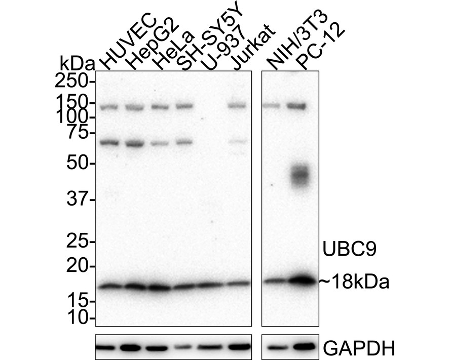
Fig1:Western blot analysis of UBC9 on different lysates with Rabbit anti-UBC9 antibody at 1/1,000 dilution. Lane 1: HUVEC cell lysate, Lane 2: HepG2 cell lysate, Lane 3: HeLa cell lysate, Lane 4: SH-SY5Y cell lysate, Lane 5: U-937 cell lysate, Lane 6: Jurkat cell lysate, Lane 7: NIH/3T3 cell lysate, Lane 8: PC-12 cell lysate, Lysates/proteins at 20 µg/Lane. Exposure time: 3 minutes; 4-20% SDS-PAGE gel. Proteins were transferred to a PVDF membrane and blocked with 5% NFDM/TBST for 1 hour at room temperature. The primary antibody at 1/1,000 dilution was used in 5% NFDM/TBST at 4℃ overnight. Goat Anti-Rabbit IgG - HRP Secondary Antibody at 1/50,000 dilution was used for 1 hour at room temperature.Application
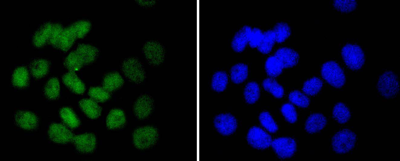
Fig2: ICC staining UBC9 in A431 cells (green). The nuclear counter stain is DAPI (blue). Cells were fixed in paraformaldehyde, permeabilised with 0.25% Triton X100/PBS.Application
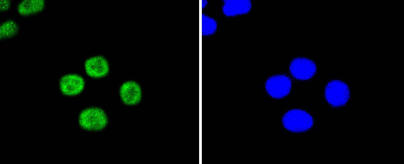
Fig3: ICC staining UBC9 in HepG2 cells (green). The nuclear counter stain is DAPI (blue). Cells were fixed in paraformaldehyde, permeabilised with 0.25% Triton X100/PBS.Application
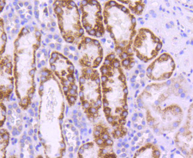
Fig4: Immunohistochemical analysis of paraffin-embedded human kidney tissue using anti-UBC9 antibody. Counter stained with hematoxylin.Application
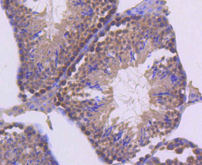
Fig5: Immunohistochemical analysis of paraffin-embedded mouse testis tissue using anti-UBC9 antibody. Counter stained with hematoxylin.| Product Name | UBC9 Recombinant Rabbit Monoclonal Antibody |
|---|---|
| Antibody Type | Primary Antibodies |
| Product description | UBC9 is a component of the ubiquitin-mediated proteolytic pathway, which targets proteins for degradation by the 26S proteasome, mediates endocytosis and directs protein subcellular localization. Ub and Ub-like molecules are systematically transferred from E2 conjugating enzymes to the targeted substrate by way of an E3 ubiquitin ligase. UBC9 functions as an E2 ubiquitin conjugating enzyme that preferentially associates with the ubiquitin homolog designated SUMO-1 or sentrin, a component of the sentrinization complex. Characteristic of the E2 family members, UBC9 contains a conserved cysteine residue that is required for the thio ester formation between Ub-like proteins and the E2 member, and it shares a conserved UBC domain. Substrates for UBC9 include transcription factors E12 and E47 and mitotic regulators RanBP2 and RanGAP1, which indicates that UBC9 may regulate various cellular processes including cell cycle progression and differentiation. |
| Immunogen | recombinant protein |
| Clonality | Monoclonal |
|---|---|
| Isotype | IgG |
| Host Species | Recombinant rabbit |
| Tested Applications | ICC/IFIHCWB |
| WB:1:500- 1:2000 ICC/IF:1:50-1:200 IHC:1:50-1:200 |
|
| Species Reactivity | HumanMouseRat |
| Concentration | 1mg/ml |
| Purification | Protein A |
| Alternative Names | C358B7.1 antibody
p18 antibody
SUMO 1 protein ligase antibody
SUMO conjugating enzyme UBC9 antibody
SUMO-conjugating enzyme UBC9 antibody
SUMO-protein ligase antibody
SUMO1 protein ligase antibody
UBC9 antibody
UBC9_HUMAN antibody
UBCE9 antibody
Ube2i antibody
Ubiquitin carrier protein 9 antibody
Ubiquitin carrier protein antibody
Ubiquitin carrier protein I antibody
Ubiquitin conjugating enzyme 9 antibody
Ubiquitin conjugating enzyme E2I (homologous to yeast UBC9) antibody
Ubiquitin conjugating enzyme E2I (UBC9 homolog yeast) antibody Ubiquitin conjugating enzyme UbcE2A antibody Ubiquitin like protein SUMO 1 conjugating enzyme antibody Ubiquitin protein ligase E2I antibody Ubiquitin-conjugating enzyme E2 I antibody Ubiquitin-protein ligase I antibody |
|---|---|
| Molecular Weight(MW) | 18 kDa |
| Cellular Localization | Nucleus, Cytoplasm. |
Application
Fig1:Western blot analysis of UBC9 on different lysates with Rabbit anti-UBC9 antibody at 1/1,000 dilution. Lane 1: HUVEC cell lysate, Lane 2: HepG2 cell lysate, Lane 3: HeLa cell lysate, Lane 4: SH-SY5Y cell lysate, Lane 5: U-937 cell lysate, Lane 6: Jurkat cell lysate, Lane 7: NIH/3T3 cell lysate, Lane 8: PC-12 cell lysate, Lysates/proteins at 20 µg/Lane. Exposure time: 3 minutes; 4-20% SDS-PAGE gel. Proteins were transferred to a PVDF membrane and blocked with 5% NFDM/TBST for 1 hour at room temperature. The primary antibody at 1/1,000 dilution was used in 5% NFDM/TBST at 4℃ overnight. Goat Anti-Rabbit IgG - HRP Secondary Antibody at 1/50,000 dilution was used for 1 hour at room temperature.
Application
Fig2: ICC staining UBC9 in A431 cells (green). The nuclear counter stain is DAPI (blue). Cells were fixed in paraformaldehyde, permeabilised with 0.25% Triton X100/PBS.
Application
Fig3: ICC staining UBC9 in HepG2 cells (green). The nuclear counter stain is DAPI (blue). Cells were fixed in paraformaldehyde, permeabilised with 0.25% Triton X100/PBS.
Application
Fig4: Immunohistochemical analysis of paraffin-embedded human kidney tissue using anti-UBC9 antibody. Counter stained with hematoxylin.
Application
Fig5: Immunohistochemical analysis of paraffin-embedded mouse testis tissue using anti-UBC9 antibody. Counter stained with hematoxylin.| Application Notes | WB:1:500- 1:2000 ICC/IF:1:50-1:200 IHC:1:50-1:200 |
|---|
| Form | Liquid |
|---|---|
| Storage Instructions | Store at +4℃ after thawing. Aliquot store at -20℃ or -80℃. Avoid repeated freeze / thaw cycles. |
| Storage Buffer | 1*TBS (pH7.4), 1%BSA, 40%Glycerol. Preservative: 0.05% Sodium Azide. |


