WB
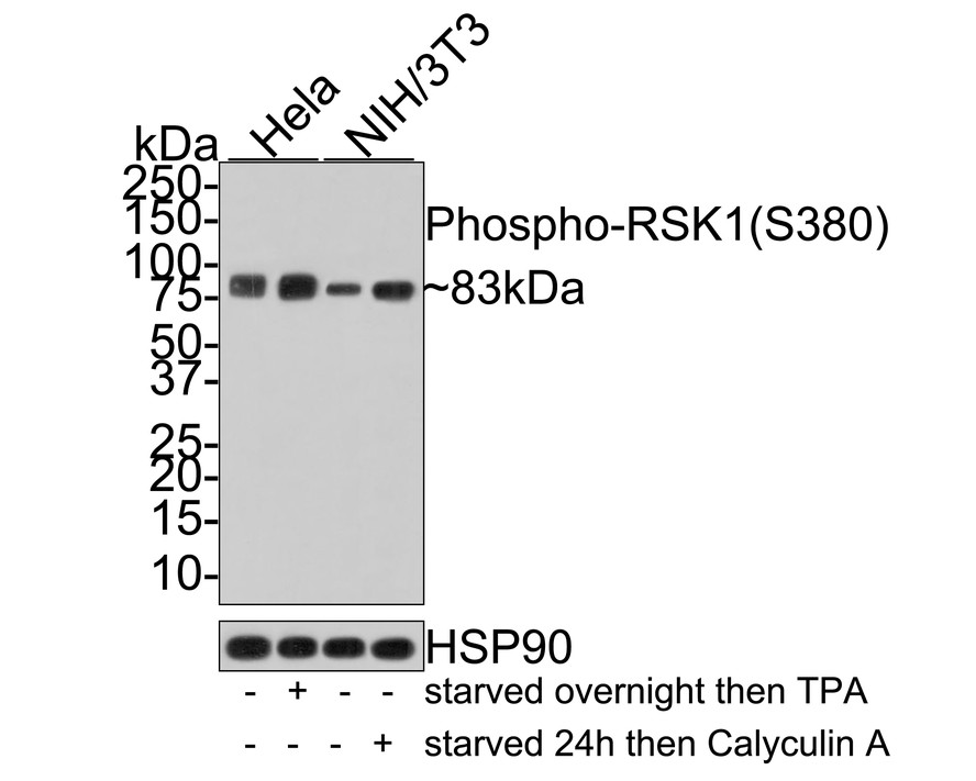
Western blot analysis of Phospho-RSK1 (S380) on different lysates with Rabbit anti-Phospho-RSK1 (S380) antibody at 1/500 dilution. Lane 1: Hela whole cell lysate, Lane 2: Hela (starved overnight) then treated with TPA (200nM 4h) cell lysate, Lane 3: NIH/3T3 whole cell lysate, Lane 4: NIH/3T3 (starved 24h) then Calyculin A (100nM 15min) cell lysate, Lysates/proteins at 10 µg/Lane. Exposure time: 1 minute; 4-20% SDS-PAGE gel. Proteins were transferred to a PVDF membrane and blocked with 5% NFDM/TBST for 1 hour at room temperature. The primary antibody at 1/500 dilution was used in 5% NFDM/TBST at room temperature for 2 hours. Goat Anti-Rabbit IgG - HRP Secondary Antibody at 1:200,000 dilution was used for 1 hour at room temperature.ICC/IF
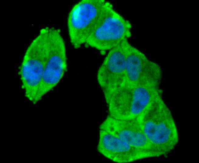
ICC staining Phospho-RSK1(S380) in Hela cells (green). The nuclear counter stain is DAPI (blue). Cells were fixed in paraformaldehyde, permeabilised with 0.25% Triton X100/PBS.ICC/IF
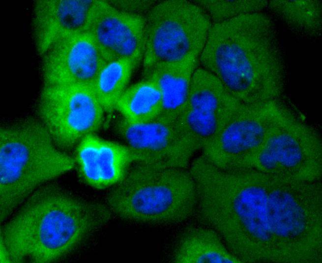
ICC staining Phospho-RSK1(S380) in A431 cells (green). The nuclear counter stain is DAPI (blue). Cells were fixed in paraformaldehyde, permeabilised with 0.25% Triton X100/PBS.ICC/IF
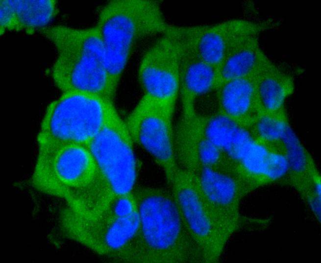
ICC staining Phospho-RSK1(S380) in 293 cells (green). The nuclear counter stain is DAPI (blue). Cells were fixed in paraformaldehyde, permeabilised with 0.25% Triton X100/PBS.| Product Name | Phospho-RSK1 (S380) Recombinant Rabbit Monoclonal Antibody |
|---|---|
| Antibody Type | Primary Antibodies |
| Product description | The family of ribosomal S6 kinases (Rsks), designated Rsk-1, Rsk-2 and Rsk-3, are important signaling intermediates that mediate responses to a broad range of ligand-activated receptor tyrosine kinases. It has been established that Rsk-3 is not activated by MAP kinase in vitro, unlike Rsk-1 and Rsk-2. A unique feature common to the three members of the Rsk family is that each possesses two non-identical complete kinase catalytic domains. The Rsk family amino-terminal kinase domain is phosphorylated on Ser 227 by 3-phosphoinositide-dependent protein kinase-1 (PDK1), which increases the kinase activity of Rsk. In the carboxy-terminal kinase domain, Rsk-1 and Rsk-2 are autophosphorylated on Ser 380 and Ser 386, respectively, which mediates the docking of PDK1 to Rsk in order to promote phosphorylation of substrates, such as histone H3. |
| Immunogen | Synthetic phospho-peptide corresponding to residues surrounding Ser380 of human RSK1. |
| Modification | p-S380 |
| Clonality | Monoclonal |
|---|---|
| Isotype | IgG |
| Host Species | Recombinant rabbit |
| Tested Applications | ICC/IFWB |
| WB:1:1000-1:5000 ICC/IF:1:50-1:200 |
|
| Species Reactivity | HumanMouseRat |
| Concentration | 1mg/ml |
| Purification | Protein A |
| Alternative Names | 90 kDa ribosomal protein S6 kinase 1 antibody
dJ590P13.1 (ribosomal protein S6 kinase 90kD polypeptide 1 antibody dJ590P13.1 antibody EC 2.7.11.1 antibody HU 1 antibody HU1 antibody KS6A1_HUMAN antibody MAP kinase activated protein kinase 1a antibody MAP kinase-activated protein kinase 1a antibody MAPK-activated protein kinase 1a antibody MAPKAP kinase 1a antibody MAPKAPK-1a antibody MAPKAPK1A antibody MGC79981 antibody Mitogen-activated protein kinase-activated protein kinase 1A antibody OTTHUMP00000004113 antibody p90 RSK1 antibody p90-RSK 1 antibody p90rsk antibody p90RSK1 antibody p90S6K antibody pp90RSK1 antibody Ribosomal protein S6 kinase 90kD 1 antibody Ribosomal protein S6 kinase 90kD polypeptide 1 antibody Ribosomal protein S6 kinase 90kDa polypeptide 1 antibody Ribosomal protein S6 kinase alpha 1 antibody Ribosomal protein S6 kinase alpha-1 antibody Ribosomal protein S6 kinase polypeptide 1 antibody Ribosomal S6 kinase 1 antibody RPS6K1 alpha antibody rps6ka antibody Rps6ka1 antibody RSK 1 antibody RSK 1 p90 antibody RSK antibody RSK-1 antibody RSK1 antibody S6K alpha 1 antibody S6K-alpha-1 antibody |
|---|---|
| Molecular Weight(MW) | 83 kDa |
| Cellular Localization | Nucleus, Cytoplasm. |
WB
Western blot analysis of Phospho-RSK1 (S380) on different lysates with Rabbit anti-Phospho-RSK1 (S380) antibody at 1/500 dilution. Lane 1: Hela whole cell lysate, Lane 2: Hela (starved overnight) then treated with TPA (200nM 4h) cell lysate, Lane 3: NIH/3T3 whole cell lysate, Lane 4: NIH/3T3 (starved 24h) then Calyculin A (100nM 15min) cell lysate, Lysates/proteins at 10 µg/Lane. Exposure time: 1 minute; 4-20% SDS-PAGE gel. Proteins were transferred to a PVDF membrane and blocked with 5% NFDM/TBST for 1 hour at room temperature. The primary antibody at 1/500 dilution was used in 5% NFDM/TBST at room temperature for 2 hours. Goat Anti-Rabbit IgG - HRP Secondary Antibody at 1:200,000 dilution was used for 1 hour at room temperature.
ICC/IF
ICC staining Phospho-RSK1(S380) in Hela cells (green). The nuclear counter stain is DAPI (blue). Cells were fixed in paraformaldehyde, permeabilised with 0.25% Triton X100/PBS.
ICC/IF
ICC staining Phospho-RSK1(S380) in A431 cells (green). The nuclear counter stain is DAPI (blue). Cells were fixed in paraformaldehyde, permeabilised with 0.25% Triton X100/PBS.
ICC/IF
ICC staining Phospho-RSK1(S380) in 293 cells (green). The nuclear counter stain is DAPI (blue). Cells were fixed in paraformaldehyde, permeabilised with 0.25% Triton X100/PBS.| Application Notes | WB:1:1000-1:5000 ICC/IF:1:50-1:200 |
|---|
| Form | Liquid |
|---|---|
| Storage Instructions | Store at +4℃ after thawing. Aliquot store at -20℃ or -80℃. Avoid repeated freeze / thaw cycles. |
| Storage Buffer | 1*TBS (pH7.4), 1%BSA, 40%Glycerol. Preservative: 0.05% Sodium Azide. |


