Application
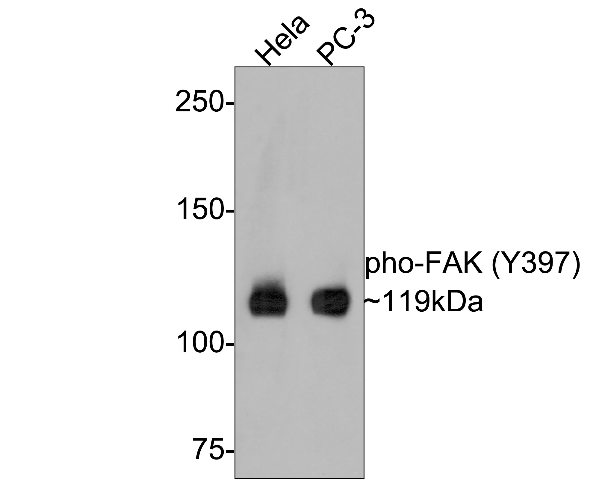
Fig1: Western blot analysis of Phospho-FAK (Y397) on different lysates with Rabbit anti-Phospho-FAK (Y397) antibody at 1/500 dilution. Lane 1: Hela cell lysate, Lane 2: PC-3 cell lysate, Lysates/proteins at 10 µg/Lane. Exposure time: 2 minutes; 6% SDS-PAGE gel. Proteins were transferred to a PVDF membrane and blocked with 5% NFDM/TBST for 1 hour at room temperature. The primary antibody at 1/500 dilution was used in 5% NFDM/TBST at room temperature for 2 hours. Goat Anti-Rabbit IgG - HRP Secondary Antibody at 1:300,000 dilution was used for 1 hour at room temperature.Application
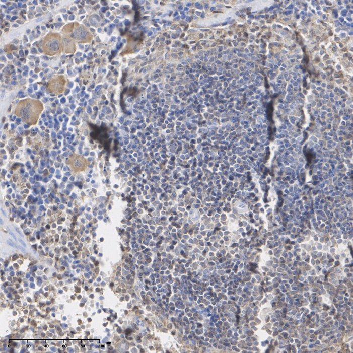
Fig2: Immunohistochemical analysis of paraffin-embedded mouse spleen tissue with Rabbit anti-Phospho-FAK (Y397) antibody at 1/200 dilution. The section was pre-treated using heat mediated antigen retrieval with Tris-EDTA buffer (pH 9.0) for 20 minutes. The tissues were blocked in 1% BSA for 20 minutes at room temperature, washed with ddH2O and PBS, and then probed with the primary antibody at 1/200 dilution for 1 hour at room temperature. The detection was performed using an HRP conjugated compact polymer system. DAB was used as the chromogen. Tissues were counterstained with hematoxylin and mounted with DPX.Application
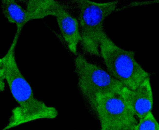
Fig3: ICC staining phospho-FAK (Y397) in NIH/3T3 cells (green). The nuclear counter stain is DAPI (blue). Cells were fixed in paraformaldehyde, permeabilised with 0.25% Triton X100/PBS.| Product Name | Phospho-FAK (Y397) Recombinant Rabbit Monoclonal Antibody |
|---|---|
| Antibody Type | Primary Antibodies |
| Product description | Activation of integrins in the extracellular matrix (ECM) of eukaryotic cells promotes the formation of membrane adhesion complexes, known as focal adhesions, which can include cytoskeletal proteins and protein tyrosine kinases, such as focal adhesion kinase (FAK). Phosphorylation events occurring within focal adhesions influence numerous processes that include mitogenic signaling, cell survival, and cell motility. FAK is a non-receptor tyrosine kinase that is ubiquitously expressed and highly conserved between species. FAK is recruited by Integrin clusters and variably phosphorylated depending on the effector molecules present in the focal adhesion. Phosphorylation of FAK Tyr 397 decreases during serum starvation, contact inhibition, and cell cycle arrest, all conditions under which activating FAK Tyr 407 phosphorylation increases. |
| Immunogen | Synthetic phospho-peptide corresponding to residues surrounding Tyr397 of human FAK. |
| Modification | p-Y397 |
| Clonality | monoclonal |
|---|---|
| Isotype | IgG |
| Host Species | Recombinant rabbit |
| Tested Applications | ICC/IFIHCWB |
| WB:1:500-1:1000 ICC/IF:1:50-1:100 IHC:1:200 |
|
| Species Reactivity | HumanMouseRat |
| Concentration | 1mg/ml |
| Purification | Protein A |
| Alternative Names | FADK 1 antibody FADK antibody FAK related non kinase polypeptide antibody FAK1 antibody FAK1_HUMAN antibody Focal adhesion kinase 1 antibody Focal adhesion Kinase antibody Focal adhesion kinase isoform FAK Del33 antibody Focal adhesion kinase related nonkinase antibody FRNK antibody p125FAK antibody pp125FAK antibody PPP1R71 antibody Protein phosphatase 1 regulatory subunit 71 antibody Protein tyrosine kinase 2 antibody Protein-tyrosine kinase 2 antibody Ptk2 antibody PTK2 protein tyrosine kinase 2 antibody |
|---|---|
| Molecular Weight(MW) | 119 kDa |
| Cellular Localization | Cytoplasm, Nucleus, Cell membrane, Cell junction. |
Application
Fig1: Western blot analysis of Phospho-FAK (Y397) on different lysates with Rabbit anti-Phospho-FAK (Y397) antibody at 1/500 dilution. Lane 1: Hela cell lysate, Lane 2: PC-3 cell lysate, Lysates/proteins at 10 µg/Lane. Exposure time: 2 minutes; 6% SDS-PAGE gel. Proteins were transferred to a PVDF membrane and blocked with 5% NFDM/TBST for 1 hour at room temperature. The primary antibody at 1/500 dilution was used in 5% NFDM/TBST at room temperature for 2 hours. Goat Anti-Rabbit IgG - HRP Secondary Antibody at 1:300,000 dilution was used for 1 hour at room temperature.
Application
Fig2: Immunohistochemical analysis of paraffin-embedded mouse spleen tissue with Rabbit anti-Phospho-FAK (Y397) antibody at 1/200 dilution. The section was pre-treated using heat mediated antigen retrieval with Tris-EDTA buffer (pH 9.0) for 20 minutes. The tissues were blocked in 1% BSA for 20 minutes at room temperature, washed with ddH2O and PBS, and then probed with the primary antibody at 1/200 dilution for 1 hour at room temperature. The detection was performed using an HRP conjugated compact polymer system. DAB was used as the chromogen. Tissues were counterstained with hematoxylin and mounted with DPX.
Application
Fig3: ICC staining phospho-FAK (Y397) in NIH/3T3 cells (green). The nuclear counter stain is DAPI (blue). Cells were fixed in paraformaldehyde, permeabilised with 0.25% Triton X100/PBS.| Application Notes | WB:1:500-1:1000 ICC/IF:1:50-1:100 IHC:1:200 |
|---|
| Form | Liquid |
|---|---|
| Storage Instructions | Store at +4℃ after thawing. Aliquot store at -20℃ or -80℃. Avoid repeated freeze / thaw cycles. |
| Storage Buffer | 1*TBS (pH7.4), 1%BSA, 40%Glycerol. Preservative: 0.05% Sodium Azide. |


