WB
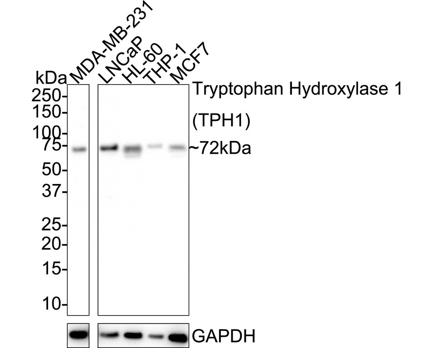
Western blot analysis of Tryptophan Hydroxylase 1 (TPH1) on different lysates with Rabbit anti-Tryptophan Hydroxylase 1 (TPH1) antibody at 1/1,000 dilution. Lane 1: MDA-MB-231 cell lysate, Lane 2: LNCaP cell lysate, Lane 3: HL-60 cell lysate, Lane 4: THP-1 cell lysate, Lane 5: MCF7 cell lysate, Lysates/proteins at 20 µg/Lane. Exposure time: 3 minutes; 4-20% SDS-PAGE gel. Proteins were transferred to a PVDF membrane and blocked with 5% NFDM/TBST for 1 hour at room temperature. The primary antibody at 1/1,000 dilution was used in 5% NFDM/TBST at 4℃ overnight. Goat Anti-Rabbit IgG - HRP Secondary Antibody at 1/50,000 dilution was used for 1 hour at room temperature.IHC
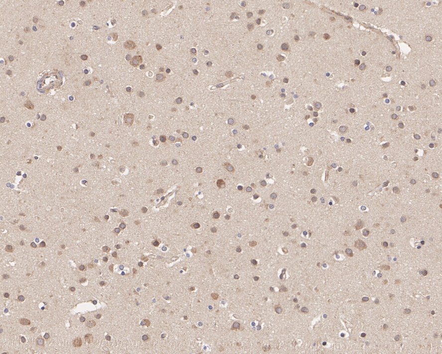
Immunohistochemical analysis of paraffin-embedded human brain tissue with Rabbit anti-Tryptophan Hydroxylase 1 (TPH1) antibody at 1/200 dilution. The section was pre-treated using heat mediated antigen retrieval with Tris-EDTA buffer (pH 9.0) for 20 minutes. The tissues were blocked in 1% BSA for 20 minutes at room temperature, washed with ddH2O and PBS, and then probed with the primary antibody at 1/200 dilution for 1 hour at room temperature. The detection was performed using an HRP conjugated compact polymer system. DAB was used as the chromogen. Tissues were counterstained with hematoxylin and mounted with DPX.IHC
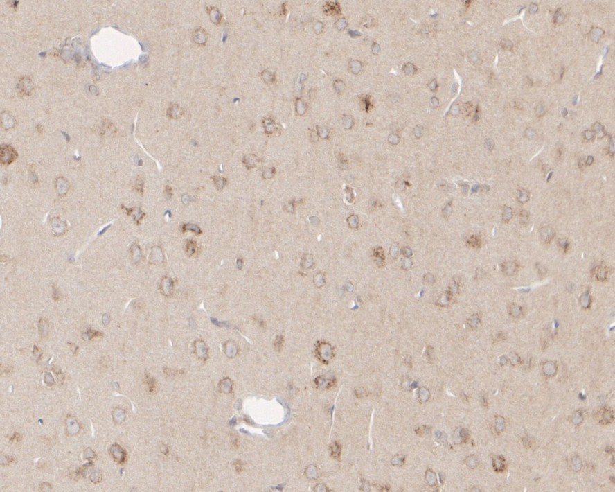
Immunohistochemical analysis of paraffin-embedded rat brain tissue with Rabbit anti-Tryptophan Hydroxylase 1 (TPH1) antibody at 1/200 dilution. The section was pre-treated using heat mediated antigen retrieval with Tris-EDTA buffer (pH 9.0) for 20 minutes. The tissues were blocked in 1% BSA for 20 minutes at room temperature, washed with ddH2O and PBS, and then probed with the primary antibody at 1/200 dilution for 1 hour at room temperature. The detection was performed using an HRP conjugated compact polymer system. DAB was used as the chromogen. Tissues were counterstained with hematoxylin and mounted with DPX.FC
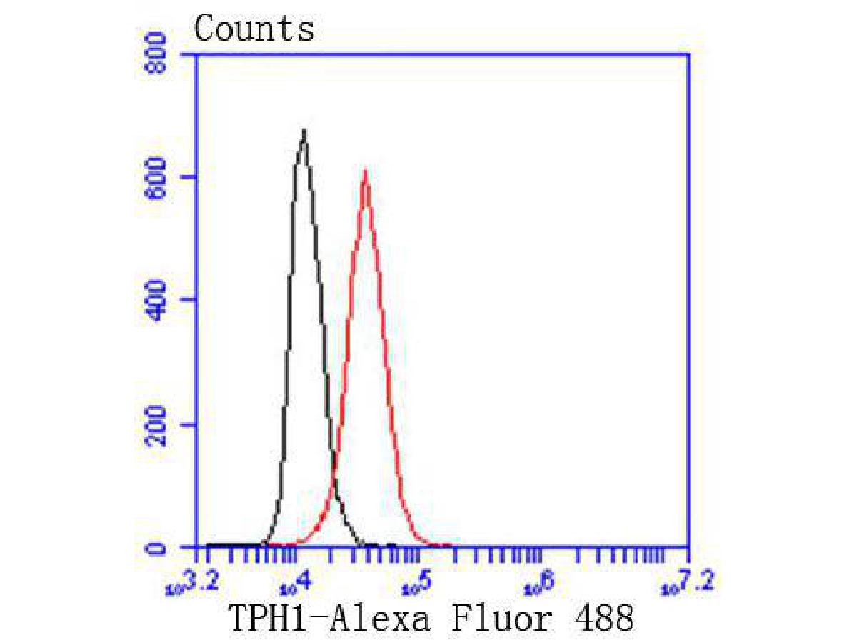
Flow cytometric analysis of Tryptophan Hydroxylase 1 (TPH1) was done on Hela cells. The cells were fixed, permeabilized and stained with the primary antibody (1/50) (red). After incubation of the primary antibody at room temperature for an hour, the cells were stained with a Alexa Fluor 488-conjugated Goat anti-Rabbit IgG Secondary antibody at 1/1000 dilution for 30 minutes.Unlabelled sample was used as a control (cells without incubation with primary antibody; black).| Product Name | Tryptophan Hydroxylase 1 (TPH1) Recombinant Rabbit Monoclonal Antibody |
|---|---|
| Antibody Type | Primary Antibodies |
| Product description | Phenylalanine hydroxylase (PAH), tyrosine hydroxylase (TH) and tryptophan hydroxylase (TPH) comprise a small family of monooxygenases that use tetrahydropterine as a cofactor during the catabolism of aromatic L-amino acids. PAH, TH and TPH all contain catalytic domains with an amino-terminal regulatory domain and a short carboxy-terminal tetramerization domain. Each of these enzymes also contains a single ferrous iron atom, which is bound to two histidines and a glutamate and is likely to be involved in the formation of the hydroxylating intermediate. TPH is the first and rate-limiting step in the biosynthesis of serotonin in the central nervous system and melatonin in the pineal gland. Alteration of TPH function may be a key factor in the pathology of several neuropsychiatric disorders associated with serotonin, including depression, aggression, alcoholism and schizophrenia. For instance, L-DOPA, which is used as a common therapy for Parkinson’s disease (PD) patients, inhibits TPH function, which subsequently, is thought to contribute to the onset of depression in PD patients. |
| Immunogen | Synthetic peptide within Human TPH1 aa 372-419 / 444. |
| Clonality | Monoclonal |
|---|---|
| Isotype | IgG |
| Host Species | Recombinant rabbit |
| Tested Applications | FCIHCWB |
| WB:1:500-1:1000 IHC:1:50-1:200 FC:1:50-1:100 |
|
| Species Reactivity | HumanMouseRat |
| Concentration | 1mg/ml |
| Purification | Protein A |
| Alternative Names | Indoleacetic acid 5 hydroxylase antibody L tryptophan hydroxylase antibody MGC119994 antibody TPH 1 antibody TPH antibody TPH1 antibody TPH1_HUMAN antibody TPRH antibody TRPH antibody Tryptophan 5 hydroxylase 1 antibody Tryptophan 5 monooxygenase 1 antibody Tryptophan 5 monooxygenase antibody Tryptophan 5-hydroxylase 1 antibody Tryptophan 5-monooxygenase 1 antibody Tryptophan hydroxylase 1 antibody |
|---|---|
| Molecular Weight(MW) | 51kDa(Observed band size: 72kDa) |
| Cellular Localization | Cytosol, neuron projection. |
WB
Western blot analysis of Tryptophan Hydroxylase 1 (TPH1) on different lysates with Rabbit anti-Tryptophan Hydroxylase 1 (TPH1) antibody at 1/1,000 dilution. Lane 1: MDA-MB-231 cell lysate, Lane 2: LNCaP cell lysate, Lane 3: HL-60 cell lysate, Lane 4: THP-1 cell lysate, Lane 5: MCF7 cell lysate, Lysates/proteins at 20 µg/Lane. Exposure time: 3 minutes; 4-20% SDS-PAGE gel. Proteins were transferred to a PVDF membrane and blocked with 5% NFDM/TBST for 1 hour at room temperature. The primary antibody at 1/1,000 dilution was used in 5% NFDM/TBST at 4℃ overnight. Goat Anti-Rabbit IgG - HRP Secondary Antibody at 1/50,000 dilution was used for 1 hour at room temperature.
IHC
Immunohistochemical analysis of paraffin-embedded human brain tissue with Rabbit anti-Tryptophan Hydroxylase 1 (TPH1) antibody at 1/200 dilution. The section was pre-treated using heat mediated antigen retrieval with Tris-EDTA buffer (pH 9.0) for 20 minutes. The tissues were blocked in 1% BSA for 20 minutes at room temperature, washed with ddH2O and PBS, and then probed with the primary antibody at 1/200 dilution for 1 hour at room temperature. The detection was performed using an HRP conjugated compact polymer system. DAB was used as the chromogen. Tissues were counterstained with hematoxylin and mounted with DPX.
IHC
Immunohistochemical analysis of paraffin-embedded rat brain tissue with Rabbit anti-Tryptophan Hydroxylase 1 (TPH1) antibody at 1/200 dilution. The section was pre-treated using heat mediated antigen retrieval with Tris-EDTA buffer (pH 9.0) for 20 minutes. The tissues were blocked in 1% BSA for 20 minutes at room temperature, washed with ddH2O and PBS, and then probed with the primary antibody at 1/200 dilution for 1 hour at room temperature. The detection was performed using an HRP conjugated compact polymer system. DAB was used as the chromogen. Tissues were counterstained with hematoxylin and mounted with DPX.
FC
Flow cytometric analysis of Tryptophan Hydroxylase 1 (TPH1) was done on Hela cells. The cells were fixed, permeabilized and stained with the primary antibody (1/50) (red). After incubation of the primary antibody at room temperature for an hour, the cells were stained with a Alexa Fluor 488-conjugated Goat anti-Rabbit IgG Secondary antibody at 1/1000 dilution for 30 minutes.Unlabelled sample was used as a control (cells without incubation with primary antibody; black).| Application Notes | WB:1:500-1:1000 IHC:1:50-1:200 FC:1:50-1:100 |
|---|
| Form | Liquid |
|---|---|
| Storage Instructions | Store at +4℃ after thawing. Aliquot store at -20℃ or -80℃. Avoid repeated freeze / thaw cycles. |
| Storage Buffer | 1*TBS (pH7.4), 1%BSA, 40%Glycerol. Preservative: 0.05% Sodium Azide. |


