WB
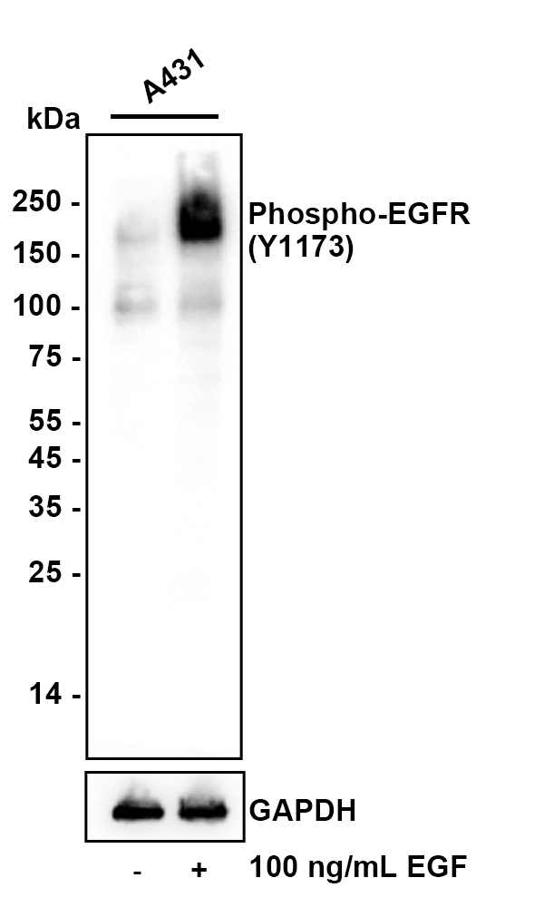
Western blot analysis of Phospho-EGFR (Y1173) on different lysates with Rabbit anti-Phospho-EGFR (Y1173) antibody at 1/2,000 dilution. Lane 1: A431 whole cell lysate, Lane 2: A431 treated with 100ng/mL EGF for 30 minutes whole cell lysate, Lysates/proteins at 10 µg/Lane. Exposure time: 2 minute; 4-20% SDS-PAGE gel. Proteins were transferred to a PVDF membrane and blocked with 5% NFDM/TBST for 1 hour at room temperature. The primary antibody at 1/2,000 dilution was used in Dilution buffer at 4℃ overnight. Goat Anti-Rabbit IgG - HRP Secondary Antibody at 1/200,000 dilution was used for 1 hour at room temperature.IHC
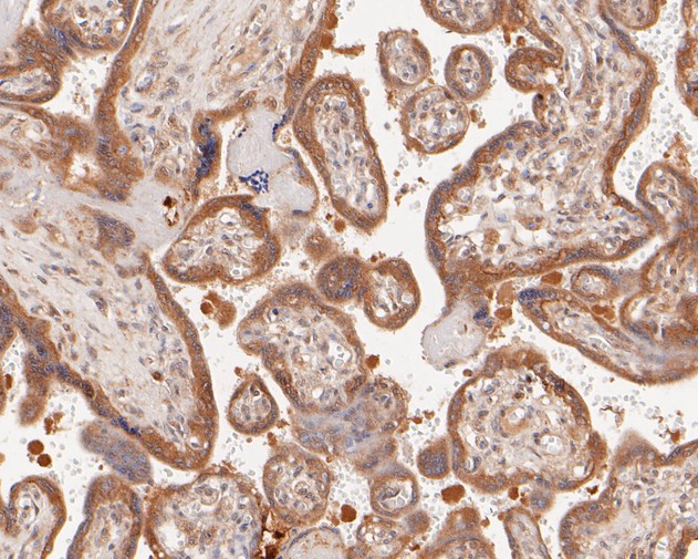
Immunohistochemical analysis of paraffin-embedded human placenta tissue using anti-Phospho-EGFR (Y1173) antibody. The section was pre-treated using heat mediated antigen retrieval with Tris-EDTA buffer (pH 8.0-8.4) for 20 minutes.The tissues were blocked in 5% BSA for 30 minutes at room temperature, washed with ddH2O and PBS, and then probed with the primary antibody (1/50) for 30 minutes at room temperature. The detection was performed using an HRP conjugated compact polymer system. DAB was used as the chromogen. Tissues were counterstained with hematoxylin and mounted with DPX.IHC
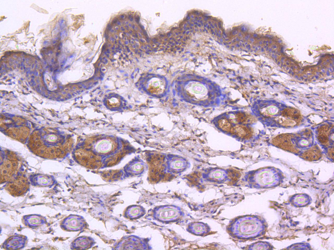
Immunohistochemical analysis of paraffin-embedded mouse skin tissue using anti- Phospho-EGFR(pY1173) antibody. Counter stained with hematoxylin.ICC/IF
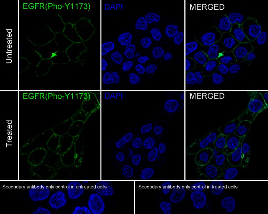
Immunocytochemistry analysis of A431 cells treated with or without 100ng/mL EGF for 5 minutes labeling Phospho-EGFR (Y1173) with Rabbit anti-Phospho-EGFR (Y1173) antibody at 1/50 dilution. Cells were fixed in 4% paraformaldehyde for 20 minutes at 37 ℃, permeabilized with 0.1% Triton X-100 in PBS permeabilization for 5 minutes, and then blocked with 2% negative goat serum for 60 minutes at room temperature. Cells were then incubated with Rabbit anti-Phospho-EGFR (Y1173) antibody at 1/50 dilution in 1% BSA overnight at 4 ℃. Goat Anti-Rabbit IgG H&L (488) was used as the secondary antibody at 1/1,000 dilution. PBS instead of the primary antibody was used as the secondary antibody only control. Nuclear DNA was labelled in blue with DAPI.| Product Name | Phospho-EGFR (Y1173) Recombinant Rabbit Monoclonal Antibody |
|---|---|
| Antibody Type | Primary Antibodies |
| Product description | Epidermal growth factor mediates its effects on cell growth through its inter-action with a cell surface glycoprotein designated the EGF receptor. Binding of EGF or TGF alpha to the EGF receptor activates tyrosine-specific protein kinase activity intrinsic to the EGF receptor. The carboxy terminal tyrosine residues on EGFR, Tyr 1068 and Tyr 1173, are the major sites of autophosphorylation, which occurs as a result of EGF binding. Once activated, EGFR mediates the binding of the phosphotyrosine binding (PTB) domain of GRB2 through direct interactions with Tyr 1068 and Tyr 1086 and through indirect interactions with Tyr 1173 in the Ras signaling pathway. Tyr 1173 of EGFR also functions as a kinase substrate. Phosphorylation of Tyr 992, Tyr 1068 and Tyr 1086 is required for conformational change in the C-terminal tail of the EGF receptor. |
| Immunogen | Synthetic phospho-peptide corresponding to residues surrounding Tyr1173 of Human EGFR. |
| Modification | p-pY1173 |
| Clonality | Monoclonal |
|---|---|
| Isotype | IgG |
| Host Species | Recombinant rabbit |
| Tested Applications | ICC/IFIHCWB |
| WB:1:1000-1:2000 IHC:1:50-1:200 ICC/IF:1:50-1:200 |
|
| Species Reactivity | HumanMouse |
| Concentration | 1mg/ml |
| Purification | Protein A |
| Alternative Names | Avian erythroblastic leukemia viral (v erb b) oncogene homolog antibody Cell growth inhibiting protein 40 antibody Cell proliferation inducing protein 61 antibody EGF R antibody EGFR antibody EGFR_HUMAN antibody Epidermal growth factor receptor (avian erythroblastic leukemia viral (v erb b) oncogene homolog) antibody Epidermal growth factor receptor (erythroblastic leukemia viral (v erb b) oncogene homolog avian) antibody Epidermal growth factor receptor antibody erb-b2 receptor tyrosine kinase 1 antibody ERBB antibody ERBB1 antibody Errp antibody HER1 antibody mENA antibody NISBD2 antibody Oncogen ERBB antibody PIG61 antibody Proto-oncogene c-ErbB-1 antibody Receptor tyrosine protein kinase ErbB 1 antibody Receptor tyrosine-protein kinase ErbB-1 antibody SA7 antibody Species antigen 7 antibody Urogastrone antibody v-erb-b Avian erythroblastic leukemia viral oncogen homolog antibody wa2 antibody Wa5 antibody |
|---|---|
| Molecular Weight(MW) | 135 kDa(Observed band size: 170 kDa) |
| Cellular Localization | Cell membrane, Nucleus membrane, Nucleus, Endoplasmic reticulum membrane, Golgi apparatus membrane. |
| SwissProt ID | P00533 |
|---|
WB
Western blot analysis of Phospho-EGFR (Y1173) on different lysates with Rabbit anti-Phospho-EGFR (Y1173) antibody at 1/2,000 dilution. Lane 1: A431 whole cell lysate, Lane 2: A431 treated with 100ng/mL EGF for 30 minutes whole cell lysate, Lysates/proteins at 10 µg/Lane. Exposure time: 2 minute; 4-20% SDS-PAGE gel. Proteins were transferred to a PVDF membrane and blocked with 5% NFDM/TBST for 1 hour at room temperature. The primary antibody at 1/2,000 dilution was used in Dilution buffer at 4℃ overnight. Goat Anti-Rabbit IgG - HRP Secondary Antibody at 1/200,000 dilution was used for 1 hour at room temperature.
IHC
Immunohistochemical analysis of paraffin-embedded human placenta tissue using anti-Phospho-EGFR (Y1173) antibody. The section was pre-treated using heat mediated antigen retrieval with Tris-EDTA buffer (pH 8.0-8.4) for 20 minutes.The tissues were blocked in 5% BSA for 30 minutes at room temperature, washed with ddH2O and PBS, and then probed with the primary antibody (1/50) for 30 minutes at room temperature. The detection was performed using an HRP conjugated compact polymer system. DAB was used as the chromogen. Tissues were counterstained with hematoxylin and mounted with DPX.
IHC
Immunohistochemical analysis of paraffin-embedded mouse skin tissue using anti- Phospho-EGFR(pY1173) antibody. Counter stained with hematoxylin.
ICC/IF
Immunocytochemistry analysis of A431 cells treated with or without 100ng/mL EGF for 5 minutes labeling Phospho-EGFR (Y1173) with Rabbit anti-Phospho-EGFR (Y1173) antibody at 1/50 dilution. Cells were fixed in 4% paraformaldehyde for 20 minutes at 37 ℃, permeabilized with 0.1% Triton X-100 in PBS permeabilization for 5 minutes, and then blocked with 2% negative goat serum for 60 minutes at room temperature. Cells were then incubated with Rabbit anti-Phospho-EGFR (Y1173) antibody at 1/50 dilution in 1% BSA overnight at 4 ℃. Goat Anti-Rabbit IgG H&L (488) was used as the secondary antibody at 1/1,000 dilution. PBS instead of the primary antibody was used as the secondary antibody only control. Nuclear DNA was labelled in blue with DAPI.| Application Notes | WB:1:1000-1:2000 IHC:1:50-1:200 ICC/IF:1:50-1:200 |
|---|
| Form | Liquid |
|---|---|
| Storage Instructions | Store at +4℃ after thawing. Aliquot store at -20℃ or -80℃. Avoid repeated freeze / thaw cycles. |
| Storage Buffer | 1*TBS (pH7.4), 1%BSA, 40%Glycerol. Preservative: 0.05% Sodium Azide. |


