WB
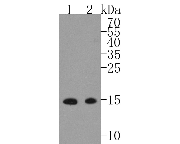
Western blot analysis of MelanA on different lysates. Proteins were transferred to a PVDF membrane and blocked with 5% BSA in PBS for 1 hour at room temperature. The primary antibody (1/500) was used in 5% BSA at room temperature for 2 hours. Goat Anti-Rabbit IgG - HRP Secondary Antibody at 1:5,000 dilution was used for 1 hour at room temperature. Positive control: Lane 1: Melanoma tissue lysate, Lane 2: B16F1 cell lysate.IHC
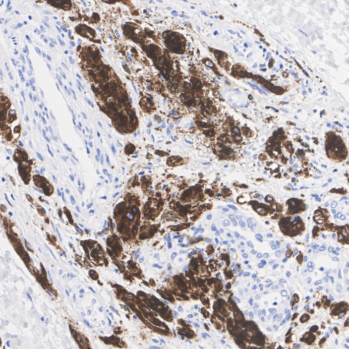
Immunohistochemical analysis of paraffin-embedded human melanoma tissue with Rabbit anti-MelanA antibody at 1/200 dilution. The section was pre-treated using heat mediated antigen retrieval with Tris-EDTA buffer (pH 9.0) for 20 minutes. The tissues were blocked in 1% BSA for 20 minutes at room temperature, washed with ddH2O and PBS, and then probed with the primary antibody at 1/200 dilution for 1 hour at room temperature. The detection was performed using an HRP conjugated compact polymer system. DAB was used as the chromogen. Tissues were counterstained with hematoxylin and mounted with DPX.IF-P
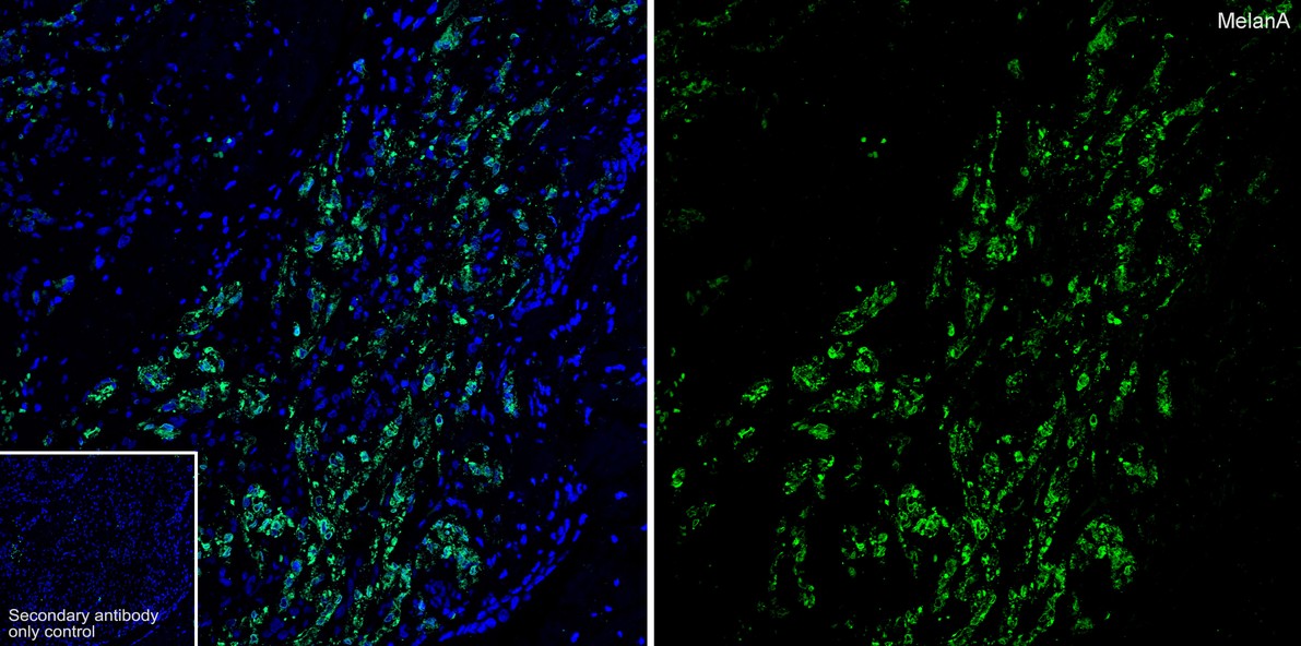
Species: Human, Site: malignant melanoma, Sample: Paraffin-embedded section.| Product Name | MelanA Recombinant Rabbit Monoclonal Antibody |
|---|---|
| Antibody Type | Primary Antibodies |
| Product description | Melanoma-associated antigens recognized by cytotoxic T lymphocytes (CTL) have been grouped into three categories: melanocyte differentiation antigens, cancer/testis-specific antigens and mutated or aberrantly expressed antigens. Many of these antigens consist of peptides that are presented to T cells by HLA molecules; they represent potential targets for cancer immunotherapy. Melan-A (also designated MART-1) is a melanocyte differentiation antigen that is specific to melanomas, melanocyte cell lines and retina. Melan-A peptide is recognized by most HLA-A2-restricted tumor-specific tumor-infiltrating lymphocytes in patients with melanoma. Antimelanoma cytotoxic T lymphocytes can be generated with a Melan-A peptide, implicating Melan-A as a potential candidate for antigen-specific immunotherapy in melanoma patients. |
| Immunogen | Synthetic peptide within Human MelanA aa 2-43 / 118. |
| Clonality | Monoclonal |
|---|---|
| Isotype | IgG |
| Host Species | Recombinant rabbit |
| Tested Applications | IF-PIHCWB |
| WB:1:500-1:2000 IHC:1:200 IF-P:1:200 |
|
| Species Reactivity | HumanMouse |
| Concentration | 1mg/ml |
| Purification | Protein A |
| Alternative Names | Antigen LB39 AA antibody Antigen LB39-AA antibody Antigen SK29 AA antibody Antigen SK29-AA antibody MAR1_HUMAN antibody MART 1 antibody MART-1 antibody MART1 antibody Melan A antibody Melan A protein antibody Melanoma antigen recognized by T cells 1 antibody Melanoma antigen recognized by T-cells 1 antibody MLAN A antibody MLANA antibody OTTHUMP00000021036 antibody OTTHUMP00000021037 antibody OTTHUMP00000021038 antibody Protein Melan-A antibody |
|---|---|
| Molecular Weight(MW) | 13kDa |
| Cellular Localization | Endoplasmic reticulum membrane, Golgi apparatus, Melanosome. |
| SwissProt ID | Q16655 |
|---|
WB
Western blot analysis of MelanA on different lysates. Proteins were transferred to a PVDF membrane and blocked with 5% BSA in PBS for 1 hour at room temperature. The primary antibody (1/500) was used in 5% BSA at room temperature for 2 hours. Goat Anti-Rabbit IgG - HRP Secondary Antibody at 1:5,000 dilution was used for 1 hour at room temperature. Positive control: Lane 1: Melanoma tissue lysate, Lane 2: B16F1 cell lysate.
IHC
Immunohistochemical analysis of paraffin-embedded human melanoma tissue with Rabbit anti-MelanA antibody at 1/200 dilution. The section was pre-treated using heat mediated antigen retrieval with Tris-EDTA buffer (pH 9.0) for 20 minutes. The tissues were blocked in 1% BSA for 20 minutes at room temperature, washed with ddH2O and PBS, and then probed with the primary antibody at 1/200 dilution for 1 hour at room temperature. The detection was performed using an HRP conjugated compact polymer system. DAB was used as the chromogen. Tissues were counterstained with hematoxylin and mounted with DPX.
IF-P
Species: Human, Site: malignant melanoma, Sample: Paraffin-embedded section.| Application Notes | WB:1:500-1:2000 IHC:1:200 IF-P:1:200 |
|---|
| Form | Liquid |
|---|---|
| Storage Instructions | Store at +4℃ after thawing. Aliquot store at -20℃ or -80℃. Avoid repeated freeze / thaw cycles. |
| Storage Buffer | 1*TBS (pH7.4), 1%BSA, 40%Glycerol. Preservative: 0.05% Sodium Azide. |


