WB
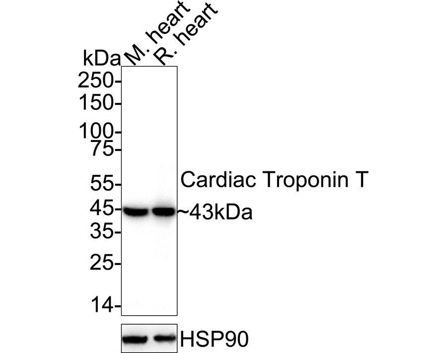
Western blot analysis of Cardiac Troponin T on different lysates with Rabbit anti-Cardiac Troponin T antibody at 1/2,000 dilution. Lane 1: Mouse heart tissue lysate, Lane 2: Rat heart tissue lysate, Lysates/proteins at 40 µg/Lane. Exposure time: 25 seconds; 4-20% SDS-PAGE gel. Proteins were transferred to a PVDF membrane and blocked with 5% NFDM/TBST for 1 hour at room temperature. The primary antibody at 1/2,000 dilution was used in 5% NFDM/TBST at 4℃ overnight. Goat Anti-Rabbit IgG - HRP Secondary Antibody at 1/50,000 dilution was used for 1 hour at room temperature.IHC
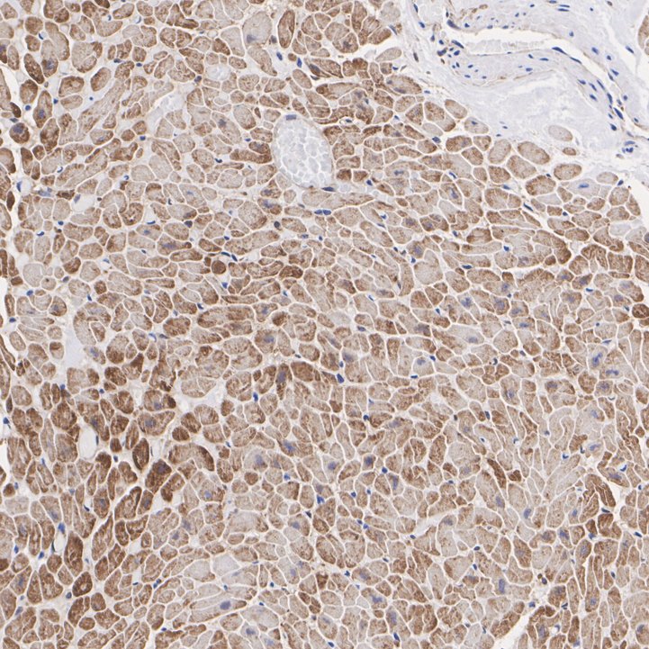
Immunohistochemical analysis of paraffin-embedded human heart tissue with Rabbit anti-Cardiac Troponin T antibody at 1/2,000 dilution. The section was pre-treated using heat mediated antigen retrieval with Tris-EDTA buffer (pH 9.0) for 20 minutes. The tissues were blocked in 1% BSA for 20 minutes at room temperature, washed with ddH2O and PBS, and then probed with the primary antibody at 1/2,000 dilution for 1 hour at room temperature. The detection was performed using an HRP conjugated compact polymer system. DAB was used as the chromogen. Tissues were counterstained with hematoxylin and mounted with DPX.IHC
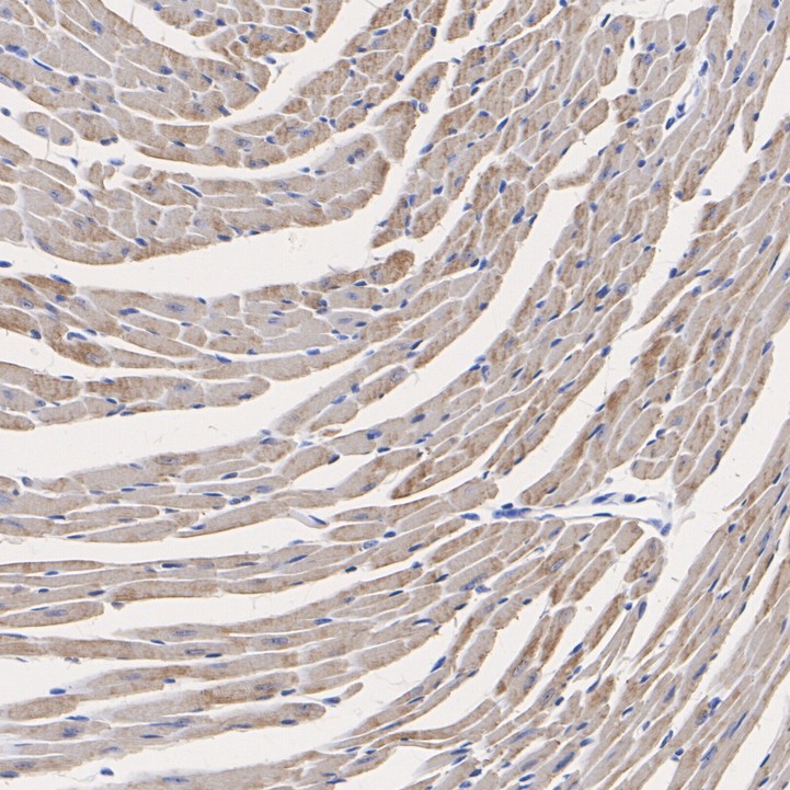
Immunohistochemical analysis of paraffin-embedded mouse heart tissue with Rabbit anti-Cardiac Troponin T antibody at 1/2,000 dilution. The section was pre-treated using heat mediated antigen retrieval with Tris-EDTA buffer (pH 9.0) for 20 minutes. The tissues were blocked in 1% BSA for 20 minutes at room temperature, washed with ddH2O and PBS, and then probed with the primary antibody at 1/2,000 dilution for 1 hour at room temperature. The detection was performed using an HRP conjugated compact polymer system. DAB was used as the chromogen. Tissues were counterstained with hematoxylin and mounted with DPX.IHC
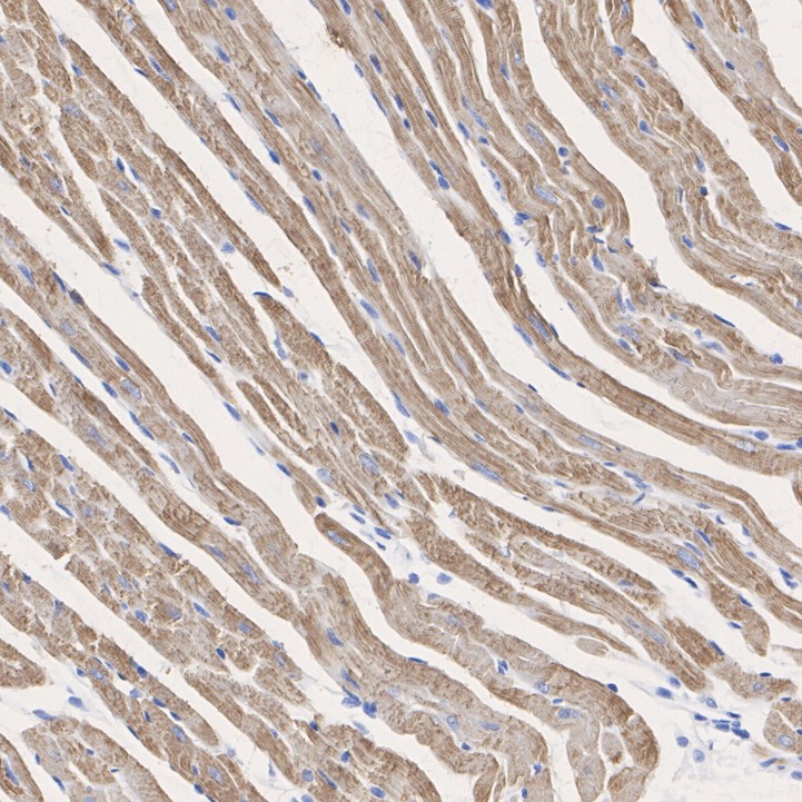
Immunohistochemical analysis of paraffin-embedded rat heart tissue with Rabbit anti-Cardiac Troponin T antibody at 1/2,000 dilution. The section was pre-treated using heat mediated antigen retrieval with Tris-EDTA buffer (pH 9.0) for 20 minutes. The tissues were blocked in 1% BSA for 20 minutes at room temperature, washed with ddH2O and PBS, and then probed with the primary antibody at 1/2,000 dilution for 1 hour at room temperature. The detection was performed using an HRP conjugated compact polymer system. DAB was used as the chromogen. Tissues were counterstained with hematoxylin and mounted with DPX.IF-P
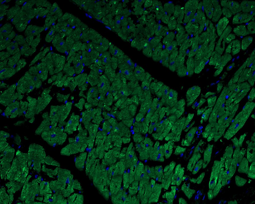
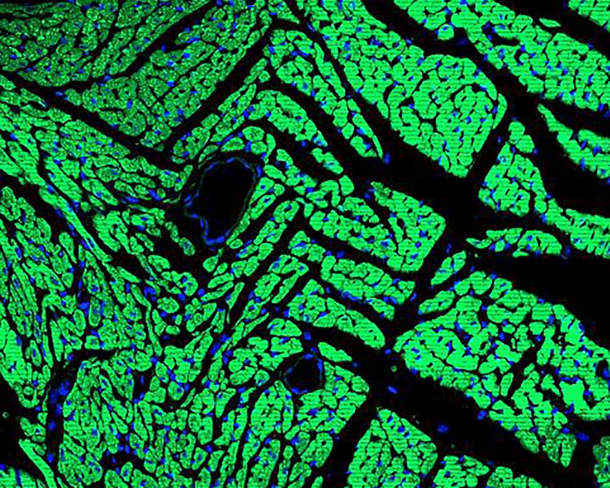
Immunofluorescence analysis of paraffin-embedded human heart tissue labeling Cardiac Troponin T with Rabbit anti-Cardiac Troponin T antibody at 1/500 dilution. The section was pre-treated using heat mediated antigen retrieval with Tris-EDTA buffer (pH 9.0) for 20 minutes. The tissues were blocked in 10% negative goat serum for 1 hour at room temperature, washed with PBS, and then probed with the primary antibody (green) at 1/200 dilution overnight at 4 ℃, washed with PBS. Goat Anti-Rabbit IgG H&L (488) was used as the secondary antibody at 1/1,000 dilution. Nuclei were counterstained with DAPI (blue).IF-P
Immunofluorescence analysis of paraffin-embedded mouse heart tissue labeling Cardiac Troponin T with Rabbit anti-Cardiac Troponin T antibody at 1/500 dilution. The section was pre-treated using heat mediated antigen retrieval with Tris-EDTA buffer (pH 9.0) for 20 minutes. The tissues were blocked in 10% negative goat serum for 1 hour at room temperature, washed with PBS, and then probed with the primary antibody ( green) at 1/200 dilution overnight at 4 ℃, washed with PBS. Goat Anti-Rabbit IgG H&L (488) was used as the secondary antibody at 1/1,000 dilution. Nuclei were counterstained with DAPI (blue).| Product Name | Cardiac Troponin T Recombinant Rabbit Monoclonal Antibody |
|---|---|
| Antibody Type | Primary Antibodies |
| Product description | Actin is a highly conserved protein that is expressed in all eukaryotic cells. Actin filaments can form both stable and labile structures and are crucial components of microvilli and the contractile apparatus of muscle cells. Myosin is a hexamer of two heavy chains (MHC) and four light chains (MLC) that interacts with Actin to generate the force for diverse cellular movements, including cytokinesis, phagocytosis and muscle contraction. Troponin facilitates the interaction between Actin and Myosin by binding to calcium. Troponin is made up of at least two subunits, which are divergent in cardiac muscle, fast skeletal muscle and slow skeletal muscle. Structures of skeletal muscle troponin are composed of Troponin C (the sensor), Troponin I (the regulator) and Troponin T (the link to the muscle thin filament). Troponin C is dumbbell-shaped and has a hydrophobic pocket that increases the contractile force of muscle fibers. Troponin C has two isoforms: fast and slow. Fast Troponin C has two calcium binding sites, while slow/cardiac Troponin C has a single calcium binding site. |
| Immunogen | Synthetic peptide within human Cardiac Troponin T aa 251-298/298. |
| Clonality | Monoclonal |
|---|---|
| Isotype | IgG |
| Host Species | Recombinant rabbit |
| Tested Applications | IF-PIHCWB |
| WB:1:2000-1:10000 IHC:1:2000 IF-P:1:500 |
|
| Species Reactivity | HumanMouseRat |
| Concentration | 1mg/ml |
| Purification | Protein A |
| Alternative Names | Cardiac muscle troponin T antibody
Cardiomyopathy dilated 1D (autosomal dominant) antibody
Cardiomyopathy hypertrophic 2 antibody
CMD1D antibody
CMH2 antibody
CMPD2 antibody
cTnT antibody
LVNC6 antibody
MGC3889 antibody
OTTHUMP00000033864 antibody
OTTHUMP00000033865 antibody
OTTHUMP00000033866 antibody
OTTHUMP00000033867 antibody
OTTHUMP00000033870 antibody
OTTHUMP00000218095 antibody
RCM3 antibody
TNNT 2 antibody
TNNT2 antibody
TNNT2_HUMAN antibody
TnTc antibody
Troponin T cardiac muscle antibody
Troponin T type 2 (cardiac) antibody
Troponin T type 2 cardiac antibody
Troponin T cardiac muscle antibody Troponin T2 cardiac antibody |
|---|---|
| Molecular Weight(MW) | 36kDa(Observed band size: 43kDa) |
| Cellular Localization | Cytoplasm, filament. |
WB
Western blot analysis of Cardiac Troponin T on different lysates with Rabbit anti-Cardiac Troponin T antibody at 1/2,000 dilution. Lane 1: Mouse heart tissue lysate, Lane 2: Rat heart tissue lysate, Lysates/proteins at 40 µg/Lane. Exposure time: 25 seconds; 4-20% SDS-PAGE gel. Proteins were transferred to a PVDF membrane and blocked with 5% NFDM/TBST for 1 hour at room temperature. The primary antibody at 1/2,000 dilution was used in 5% NFDM/TBST at 4℃ overnight. Goat Anti-Rabbit IgG - HRP Secondary Antibody at 1/50,000 dilution was used for 1 hour at room temperature.
IHC
Immunohistochemical analysis of paraffin-embedded human heart tissue with Rabbit anti-Cardiac Troponin T antibody at 1/2,000 dilution. The section was pre-treated using heat mediated antigen retrieval with Tris-EDTA buffer (pH 9.0) for 20 minutes. The tissues were blocked in 1% BSA for 20 minutes at room temperature, washed with ddH2O and PBS, and then probed with the primary antibody at 1/2,000 dilution for 1 hour at room temperature. The detection was performed using an HRP conjugated compact polymer system. DAB was used as the chromogen. Tissues were counterstained with hematoxylin and mounted with DPX.
IHC
Immunohistochemical analysis of paraffin-embedded mouse heart tissue with Rabbit anti-Cardiac Troponin T antibody at 1/2,000 dilution. The section was pre-treated using heat mediated antigen retrieval with Tris-EDTA buffer (pH 9.0) for 20 minutes. The tissues were blocked in 1% BSA for 20 minutes at room temperature, washed with ddH2O and PBS, and then probed with the primary antibody at 1/2,000 dilution for 1 hour at room temperature. The detection was performed using an HRP conjugated compact polymer system. DAB was used as the chromogen. Tissues were counterstained with hematoxylin and mounted with DPX.
IHC
Immunohistochemical analysis of paraffin-embedded rat heart tissue with Rabbit anti-Cardiac Troponin T antibody at 1/2,000 dilution. The section was pre-treated using heat mediated antigen retrieval with Tris-EDTA buffer (pH 9.0) for 20 minutes. The tissues were blocked in 1% BSA for 20 minutes at room temperature, washed with ddH2O and PBS, and then probed with the primary antibody at 1/2,000 dilution for 1 hour at room temperature. The detection was performed using an HRP conjugated compact polymer system. DAB was used as the chromogen. Tissues were counterstained with hematoxylin and mounted with DPX.
IF-P
Immunofluorescence analysis of paraffin-embedded human heart tissue labeling Cardiac Troponin T with Rabbit anti-Cardiac Troponin T antibody at 1/500 dilution. The section was pre-treated using heat mediated antigen retrieval with Tris-EDTA buffer (pH 9.0) for 20 minutes. The tissues were blocked in 10% negative goat serum for 1 hour at room temperature, washed with PBS, and then probed with the primary antibody (green) at 1/200 dilution overnight at 4 ℃, washed with PBS. Goat Anti-Rabbit IgG H&L (488) was used as the secondary antibody at 1/1,000 dilution. Nuclei were counterstained with DAPI (blue).
IF-P
Immunofluorescence analysis of paraffin-embedded mouse heart tissue labeling Cardiac Troponin T with Rabbit anti-Cardiac Troponin T antibody at 1/500 dilution. The section was pre-treated using heat mediated antigen retrieval with Tris-EDTA buffer (pH 9.0) for 20 minutes. The tissues were blocked in 10% negative goat serum for 1 hour at room temperature, washed with PBS, and then probed with the primary antibody ( green) at 1/200 dilution overnight at 4 ℃, washed with PBS. Goat Anti-Rabbit IgG H&L (488) was used as the secondary antibody at 1/1,000 dilution. Nuclei were counterstained with DAPI (blue).| Application Notes | WB:1:2000-1:10000 IHC:1:2000 IF-P:1:500 |
|---|
| Form | Liquid |
|---|---|
| Storage Instructions | Store at +4℃ after thawing. Aliquot store at -20℃ or -80℃. Avoid repeated freeze / thaw cycles. |
| Storage Buffer | 1*TBS (pH7.4), 1%BSA, 40%Glycerol. Preservative: 0.05% Sodium Azide. |


