WB
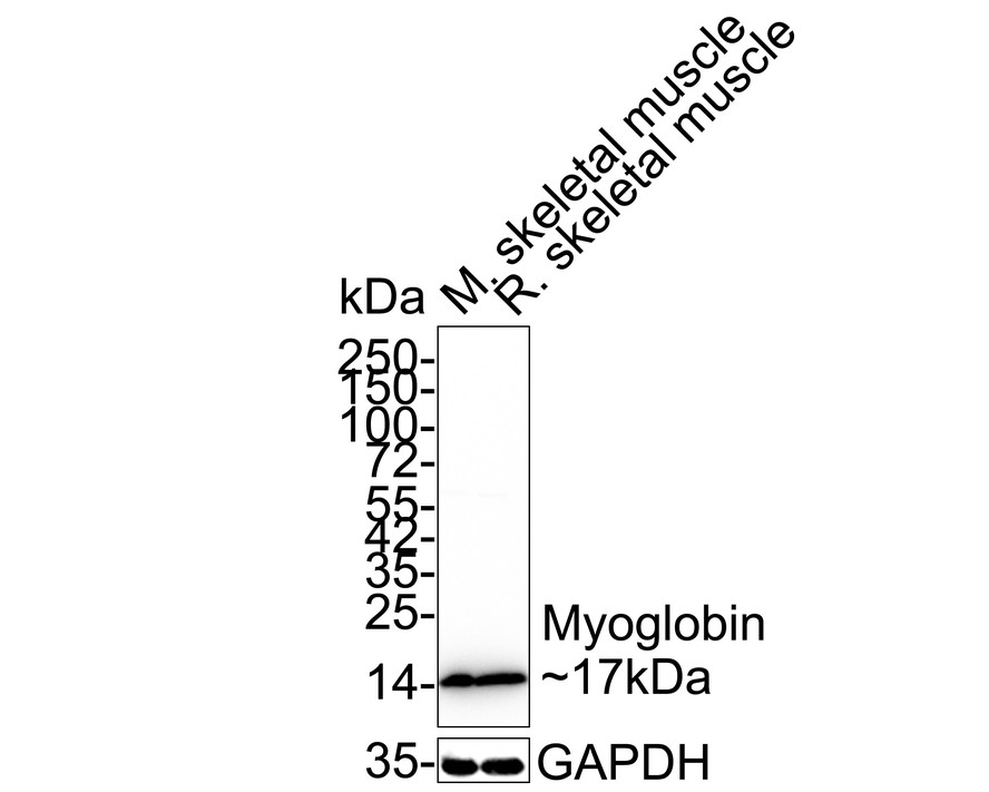
Western blot analysis of Myoglobin on different lysates with Rabbit anti-Myoglobin antibody at 1/1,000 dilution. Lane 1: Mouse skeletal muscle tissue lysate, Lane 2: Rat skeletal muscle tissue lysate, Lysates/proteins at 40 µg/Lane. Exposure time: 2 seconds; 4-20% SDS-PAGE gel. Proteins were transferred to a PVDF membrane and blocked with 5% NFDM/TBST for 1 hour at room temperature. The primary antibody at 1/1,000 dilution was used in 5% NFDM/TBST at 4℃ overnight. Goat Anti-Rabbit IgG - HRP Secondary Antibody at 1/50,000 dilution was used for 1 hour at room temperature.WB
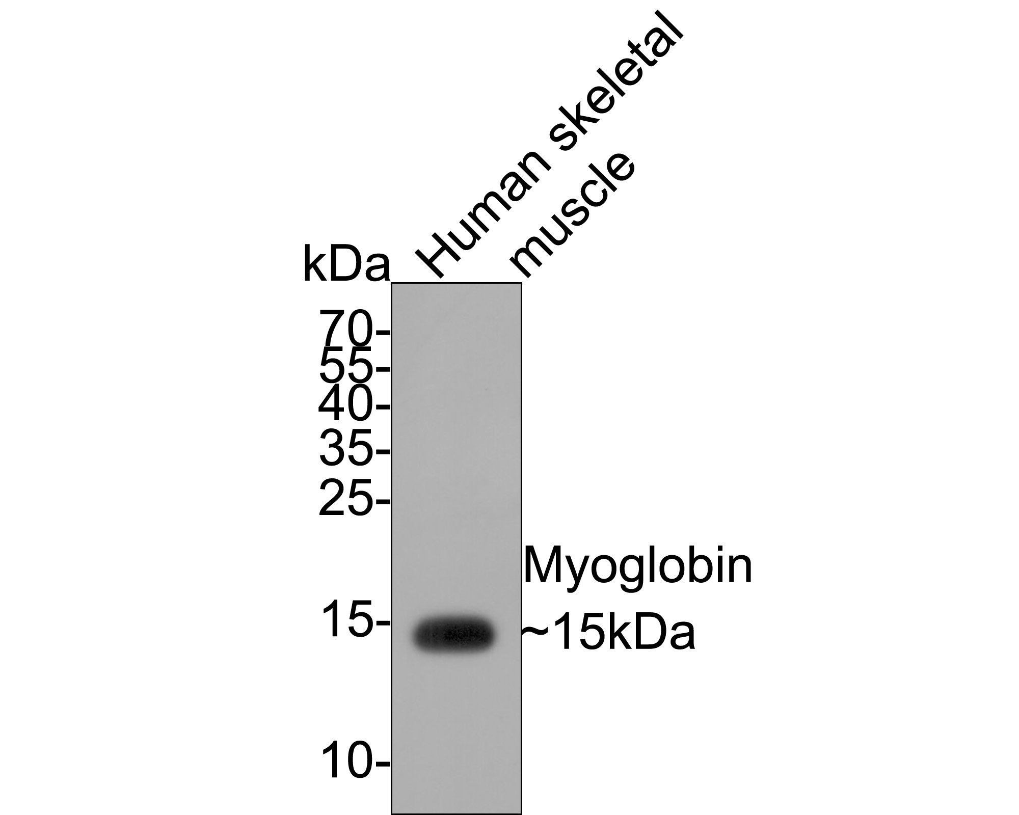
Western blot analysis of Myoglobin on human skeletal muscle tissue lysates with Rabbit anti-Myoglobin antibody at 1/500 dilution. Lysates/proteins at 10 µg/Lane. Exposure time: 1 minute; 15% SDS-PAGE gel. Proteins were transferred to a PVDF membrane and blocked with 5% NFDM/TBST for 1 hour at room temperature. The primary antibody at 1/500 dilution was used in 5% NFDM/TBST at room temperature for 2 hours. Goat Anti-Rabbit IgG - HRP Secondary Antibody at 1:300,000 dilution was used for 1 hour at room temperature.IHC
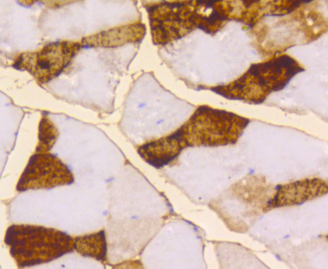
Immunohistochemical analysis of paraffin-embedded mouse skeletal muscle tissue using anti-Myoglobin antibody. Counter stained with hematoxylin.IHC
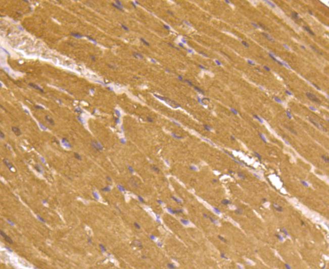
Immunohistochemical analysis of paraffin-embedded mouse heart tissue using anti-Myoglobin antibody. Counter stained with hematoxylin.ICC/IF
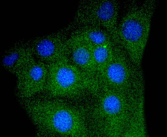
ICC staining of Myoglobin in C2C12 cells (green). Formalin fixed cells were permeabilized with 0.1% Triton X-100 in TBS for 10 minutes at room temperature and blocked with 1% Blocker BSA for 15 minutes at room temperature. Cells were probed with the primary antibody (1/50) for 1 hour at room temperature, washed with PBS. Alexa Fluor®488 Goat anti-Rabbit IgG was used as the secondary antibody at 1/1,000 dilution. The nuclear counter stain is DAPI (blue).| Product Name | Myoglobin Recombinant Rabbit Monoclonal Antibody |
|---|---|
| Antibody Type | Primary Antibodies |
| Product description | Myoglobin is a cytosolic oxygen binding protein responsible for the storage and diffusion of oxygen within myocytes. Expression of myoglobin is highest in skeletal and cardiac muscle. Myoglobin is necessary for the maintenance of mitochondrial respiration during heavy and sustained contractile activity, and it is thought to transport oxygen from erythroyctes to mitochondria. The genomic structure of myoglobin appears to be conserved across a broad range of species, and contains a putative polyadenylation signal and a polypyrimidine-rich region. Human myoglobin is specified by a single gene, and it has been identified in human smooth muscle. |
| Immunogen | Synthetic peptide within Human Myoglobin aa 121-154 / 154. |
| Clonality | Monoclonal |
|---|---|
| Isotype | IgG |
| Host Species | Recombinant rabbit |
| Tested Applications | ICC/IFIHCWB |
| WB:1:1000-1:5000 ICC/IF:1:50-1:200 IHC:1:50-1:200 |
|
| Species Reactivity | HumanMouseRat |
| Concentration | 1mg/ml |
| Purification | Protein A |
| Alternative Names | MB antibody MGC13548 antibody MYG_HUMAN antibody Myoglobin antibody PVALB antibody |
|---|---|
| Molecular Weight(MW) | 17 kDa |
| Cellular Localization | Cytoplasm. |
WB
Western blot analysis of Myoglobin on different lysates with Rabbit anti-Myoglobin antibody at 1/1,000 dilution. Lane 1: Mouse skeletal muscle tissue lysate, Lane 2: Rat skeletal muscle tissue lysate, Lysates/proteins at 40 µg/Lane. Exposure time: 2 seconds; 4-20% SDS-PAGE gel. Proteins were transferred to a PVDF membrane and blocked with 5% NFDM/TBST for 1 hour at room temperature. The primary antibody at 1/1,000 dilution was used in 5% NFDM/TBST at 4℃ overnight. Goat Anti-Rabbit IgG - HRP Secondary Antibody at 1/50,000 dilution was used for 1 hour at room temperature.
WB
Western blot analysis of Myoglobin on human skeletal muscle tissue lysates with Rabbit anti-Myoglobin antibody at 1/500 dilution. Lysates/proteins at 10 µg/Lane. Exposure time: 1 minute; 15% SDS-PAGE gel. Proteins were transferred to a PVDF membrane and blocked with 5% NFDM/TBST for 1 hour at room temperature. The primary antibody at 1/500 dilution was used in 5% NFDM/TBST at room temperature for 2 hours. Goat Anti-Rabbit IgG - HRP Secondary Antibody at 1:300,000 dilution was used for 1 hour at room temperature.
IHC
Immunohistochemical analysis of paraffin-embedded mouse skeletal muscle tissue using anti-Myoglobin antibody. Counter stained with hematoxylin.
IHC
Immunohistochemical analysis of paraffin-embedded mouse heart tissue using anti-Myoglobin antibody. Counter stained with hematoxylin.
ICC/IF
ICC staining of Myoglobin in C2C12 cells (green). Formalin fixed cells were permeabilized with 0.1% Triton X-100 in TBS for 10 minutes at room temperature and blocked with 1% Blocker BSA for 15 minutes at room temperature. Cells were probed with the primary antibody (1/50) for 1 hour at room temperature, washed with PBS. Alexa Fluor®488 Goat anti-Rabbit IgG was used as the secondary antibody at 1/1,000 dilution. The nuclear counter stain is DAPI (blue).| Application Notes | WB:1:1000-1:5000 ICC/IF:1:50-1:200 IHC:1:50-1:200 |
|---|
| Form | Liquid |
|---|---|
| Storage Instructions | Store at +4℃ after thawing. Aliquot store at -20℃ or -80℃. Avoid repeated freeze / thaw cycles. |
| Storage Buffer | 1*TBS (pH7.4), 1%BSA, 40%Glycerol. Preservative: 0.05% Sodium Azide. |


