WB
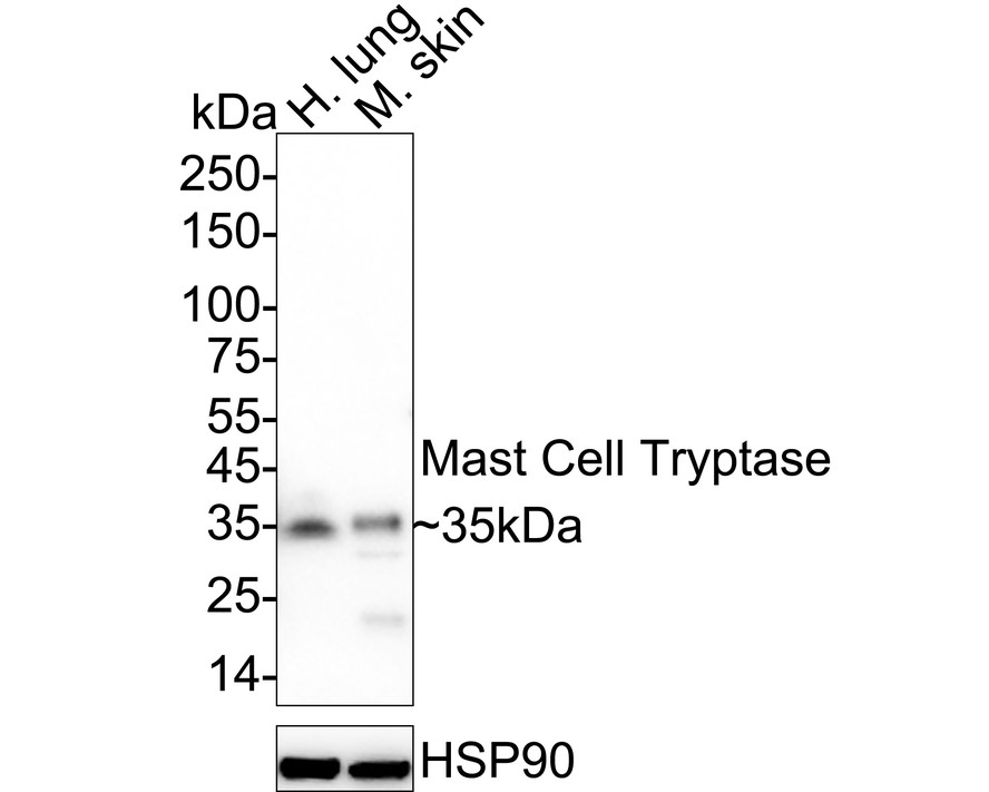
Western blot analysis of Mast Cell Tryptase on different lysates with Rabbit anti-Mast Cell Tryptase antibody at 1/1,000 dilution. Lane 1: Human lung tissue lysate (40 µg/Lane), Lane 2: Mouse skin tissue lysate (40 µg/Lane), Exposure time: 59 seconds; 4-20% SDS-PAGE gel. Proteins were transferred to a PVDF membrane and blocked with 5% NFDM/TBST for 1 hour at room temperature. The primary antibody at 1/1,000 dilution was used in 5% NFDM/TBST at 4℃ overnight. Goat Anti-Rabbit IgG - HRP Secondary Antibody at 1/50,000 dilution was used for 1 hour at room temperature.IHC
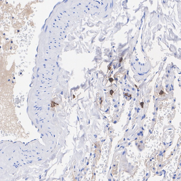
Immunohistochemical analysis of paraffin-embedded human lung tissue with Rabbit anti-Mast Cell Tryptase antibody at 1/10,000 dilution. The section was pre-treated using heat mediated antigen retrieval with Tris-EDTA buffer (pH 9.0) for 20 minutes. The tissues were blocked in 1% BSA for 20 minutes at room temperature, washed with ddH2O and PBS, and then probed with the primary antibody at 1/10,000 dilution for 1 hour at room temperature. The detection was performed using an HRP conjugated compact polymer system. DAB was used as the chromogen. Tissues were counterstained with hematoxylin and mounted with DPX.IF-P
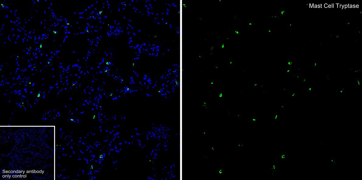
Species: Human, Tissue: Lung, Sample: Paraffin-embedded section, Antigen retrieval: Heat-mediated, Tris-EDTA buffer (pH 9.0), 20 minutes at 95℃. Wash buffer: 1× TBST, Blocking: 10% normal goat serum + 1% Triton X-100 + 0.3 M Glycine in TBST, 30 minutes at room temperature. Primary antibody: 1/1,000, overnight at 4℃. Secondary antibody: Goat Anti-Rabbit IgG (488), 1.5 hours at room temperature.mIHC
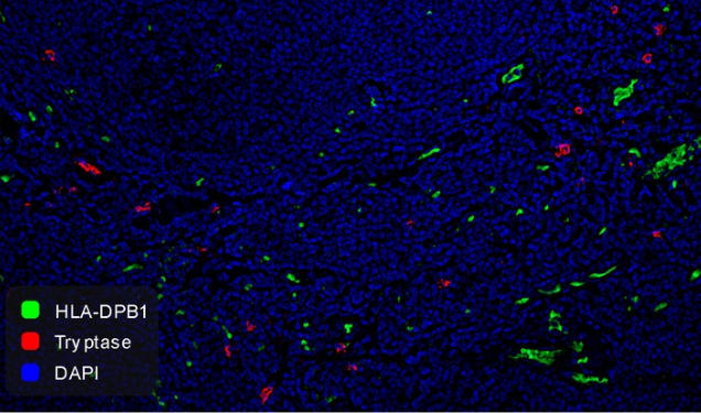
Fluorescence multiplex immunohistochemical analysis of human tonsil (Formalin/PFA-fixed paraffin-embedded sections). Panel A: the merged image of anti-HLA-DBP1 (Green) and anti-Tryptase (Red) on tonsil. HRP Conjugated UltraPolymer Goat Polyclonal Antibody HA1119/HA1120 was used as a secondary antibody. The immunostaining was performed with the Sequential Immuno-staining Kit. The section was incubated in two rounds of staining: in the order of anti-HLA-DBP1 (1/2,000 dilution) and anti-Tryptase (1/5,000 dilution) for 20 mins at room temperature. Each round was followed by a separate fluorescent tyramide signal amplification system. Heat mediated antigen retrieval with Tris-EDTA buffer (pH 9.0) for 30 mins at 95℃. DAPI (blue) was used as a nuclear counter stain. Image acquisition was performed with Zeiss Observer 7 Inverted Fluorescence Microscope.| Product Name | Mast Cell Tryptase Recombinant Rabbit Monoclonal Antibody |
|---|---|
| Antibody Type | Primary Antibodies |
| Product description | Mast cells are connective tissue cells derived from blood-forming tissues that line arterial walls and secrete substances, which mediate inflammatory and immune responses. Mast cell chymase, known as CMA1, is a major secreted serine protease that is involved in vasoactive peptide generation, extracellular matrix degradation and regulation of gland secretion. The human chymase gene, which maps to human chromosome 14q11.2, encodes a preproenzyme with a 19-amino acid signal peptide, an acidic 2-amino acid propeptide and a 226-amino acid catalytic domain. Tryptases comprise a family of trypsin-like serine proteases that are enzymatically active as heparin-stabilized tetramers. There are four functional genes for tryptase: α I, β I, β II and γ I, which map to human chromosome 16p13.3, with β tryptases representing the main isoenzymes expressed in mast cells. Mast cell proteases are a family of rodent protein homologs to human tryptases that are specifically expressed in mast cells and may serve as highly specific markers in the analysis of mast cell heterogeneity, differentiation and function. |
| Immunogen | Recombinant protein within Human Mast Cell Tryptase aa 10-275 / 275. |
| Clonality | Monoclonal |
|---|---|
| Isotype | IgG |
| Host Species | Recombinant rabbit |
| Tested Applications | IF-PIHCWBmIHC |
| WB:1:1000 IHC:1:5000-1:10000 IF-P:1:1000 mIHC:1:3000-1:5000 |
|
| Species Reactivity | HumanMouse |
| Concentration | 1mg/ml |
| Purification | Protein A |
| Alternative Names | alpha II antibody Lung tryptase antibody Mast cell alpha II tryptase antibody Mast cell beta I tryptase antibody Mast cell protease 7 antibody Mast cell protease II antibody MCP 7 antibody Pituitary tryptase antibody Skin tryptase antibody TPS 1 antibody TPS1 antibody TPS2 antibody TPSAB1 antibody TPSAB1 protein antibody TPSB1 antibody Tryptase 1 antibody Tryptase alpha 1 antibody tryptase alpha I included antibody Tryptase alpha II antibody tryptase alpha II included antibody tryptase alpha included antibody tryptase alpha/beta 1 antibody Tryptase beta 1 antibody tryptase beta I included antibody Tryptase I antibody tryptase I included antibody Tryptase III antibody Tryptase skin antibody |
|---|---|
| Molecular Weight(MW) | 30 kDa(Observed band size: 35 kDa) |
| Cellular Localization | Secreted. |
WB
Western blot analysis of Mast Cell Tryptase on different lysates with Rabbit anti-Mast Cell Tryptase antibody at 1/1,000 dilution. Lane 1: Human lung tissue lysate (40 µg/Lane), Lane 2: Mouse skin tissue lysate (40 µg/Lane), Exposure time: 59 seconds; 4-20% SDS-PAGE gel. Proteins were transferred to a PVDF membrane and blocked with 5% NFDM/TBST for 1 hour at room temperature. The primary antibody at 1/1,000 dilution was used in 5% NFDM/TBST at 4℃ overnight. Goat Anti-Rabbit IgG - HRP Secondary Antibody at 1/50,000 dilution was used for 1 hour at room temperature.
IHC
Immunohistochemical analysis of paraffin-embedded human lung tissue with Rabbit anti-Mast Cell Tryptase antibody at 1/10,000 dilution. The section was pre-treated using heat mediated antigen retrieval with Tris-EDTA buffer (pH 9.0) for 20 minutes. The tissues were blocked in 1% BSA for 20 minutes at room temperature, washed with ddH2O and PBS, and then probed with the primary antibody at 1/10,000 dilution for 1 hour at room temperature. The detection was performed using an HRP conjugated compact polymer system. DAB was used as the chromogen. Tissues were counterstained with hematoxylin and mounted with DPX.
IF-P
Species: Human, Tissue: Lung, Sample: Paraffin-embedded section, Antigen retrieval: Heat-mediated, Tris-EDTA buffer (pH 9.0), 20 minutes at 95℃. Wash buffer: 1× TBST, Blocking: 10% normal goat serum + 1% Triton X-100 + 0.3 M Glycine in TBST, 30 minutes at room temperature. Primary antibody: 1/1,000, overnight at 4℃. Secondary antibody: Goat Anti-Rabbit IgG (488), 1.5 hours at room temperature.
mIHC
Fluorescence multiplex immunohistochemical analysis of human tonsil (Formalin/PFA-fixed paraffin-embedded sections). Panel A: the merged image of anti-HLA-DBP1 (Green) and anti-Tryptase (Red) on tonsil. HRP Conjugated UltraPolymer Goat Polyclonal Antibody HA1119/HA1120 was used as a secondary antibody. The immunostaining was performed with the Sequential Immuno-staining Kit. The section was incubated in two rounds of staining: in the order of anti-HLA-DBP1 (1/2,000 dilution) and anti-Tryptase (1/5,000 dilution) for 20 mins at room temperature. Each round was followed by a separate fluorescent tyramide signal amplification system. Heat mediated antigen retrieval with Tris-EDTA buffer (pH 9.0) for 30 mins at 95℃. DAPI (blue) was used as a nuclear counter stain. Image acquisition was performed with Zeiss Observer 7 Inverted Fluorescence Microscope.| Application Notes | WB:1:1000 IHC:1:5000-1:10000 IF-P:1:1000 mIHC:1:3000-1:5000 |
|---|
| Form | Liquid |
|---|---|
| Storage Instructions | Store at +4℃ after thawing. Aliquot store at -20℃ or -80℃. Avoid repeated freeze / thaw cycles. |
| Storage Buffer | 1*TBS (pH7.4), 1%BSA, 40%Glycerol. Preservative: 0.05% Sodium Azide. |


