WB
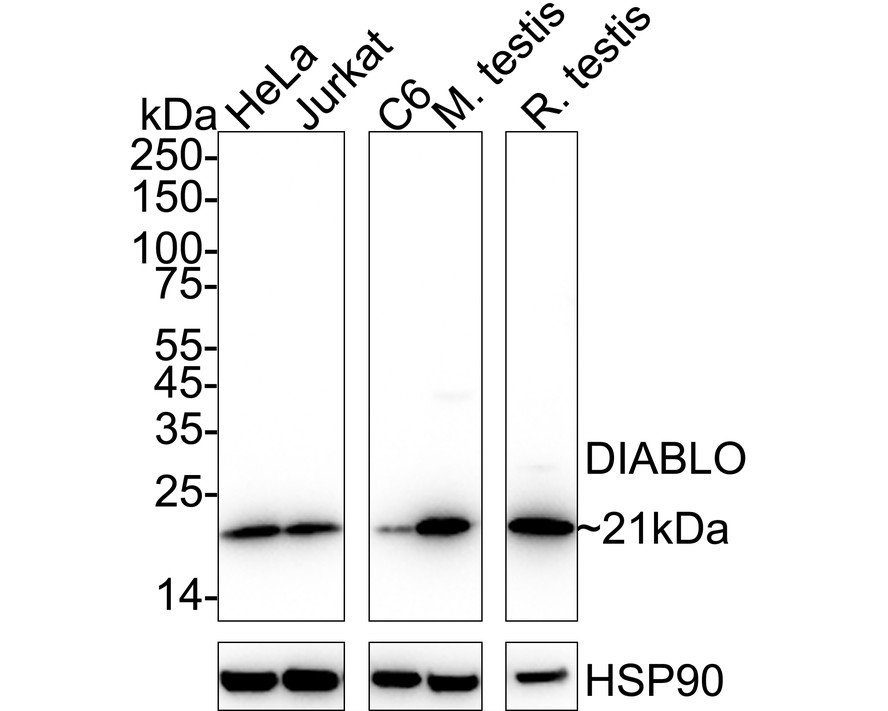
Western blot analysis of DIABLO on different lysates with Rabbit anti-DIABLO antibody at 1/2,000 dilution. Lane 1: HeLa cell lysate (20 µg/Lane), Lane 2: Jurkat cell lysate (20 µg/Lane), Lane 3: C6 cell lysate (20 µg/Lane), Lane 4: Mouse testis tissue lysate (40 µg/Lane), Lane 5: Rat testis tissue lysate (40 µg/Lane), Exposure time: 2 seconds; 4-20% SDS-PAGE gel. Proteins were transferred to a PVDF membrane and blocked with 5% NFDM/TBST for 1 hour at room temperature. The primary antibody at 1/2,000 dilution was used in 5% NFDM/TBST at 4℃ overnight. Goat Anti-Rabbit IgG - HRP Secondary Antibody at 1/50,000 dilution was used for 1 hour at room temperature.ICC/IF
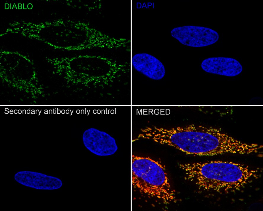
Immunocytochemistry analysis of HeLa cells labeling DIABLO with Rabbit anti-DIABLO antibody at 1/100 dilution. Cells were fixed in 4% paraformaldehyde for 15 minutes at room temperature, permeabilized with 0.1% Triton X-100 in PBS for 15 minutes at room temperature, then blocked with 1% BSA in 10% negative goat serum for 1 hour at room temperature. Cells were then incubated with Rabbit anti-DIABLO antibody at 1/100 dilution in 1% BSA in PBST overnight at 4 ℃. Goat Anti-Rabbit IgG H&L (488) was used as the secondary antibody at 1/1,000 dilution. PBS instead of the primary antibody was used as the secondary antibody only control. Counterstained with Mitotracker. Nuclear DNA was labelled in blue with DAPI.ICC/IF
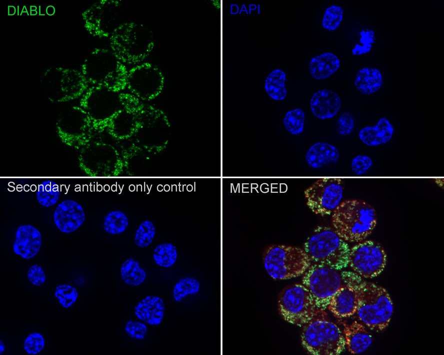
Immunocytochemistry analysis of Neuro-2a cells labeling DIABLO with Rabbit anti-DIABLO antibody at 1/100 dilution. Cells were fixed in 4% paraformaldehyde for 15 minutes at room temperature, permeabilized with 0.1% Triton X-100 in PBS for 15 minutes at room temperature, then blocked with 1% BSA in 10% negative goat serum for 1 hour at room temperature. Cells were then incubated with Rabbit anti-DIABLO antibody at 1/100 dilution in 1% BSA in PBST overnight at 4 ℃. Goat Anti-Rabbit IgG H&L (488) was used as the secondary antibody at 1/1,000 dilution. PBS instead of the primary antibody was used as the secondary antibody only control. Counterstained with Mitotracker. Nuclear DNA was labelled in blue with DAPI.ICC/IF
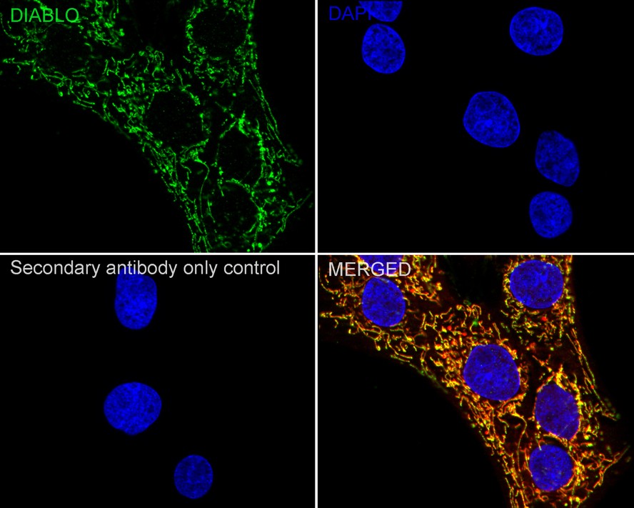
Immunocytochemistry analysis of C6 cells labeling DIABLO with Rabbit anti-DIABLO antibody at 1/100 dilution. Cells were fixed in 4% paraformaldehyde for 15 minutes at room temperature, permeabilized with 0.1% Triton X-100 in PBS for 15 minutes at room temperature, then blocked with 1% BSA in 10% negative goat serum for 1 hour at room temperature. Cells were then incubated with Rabbit anti-DIABLO antibody at 1/100 dilution in 1% BSA in PBST overnight at 4 ℃. Goat Anti-Rabbit IgG H&L (488) was used as the secondary antibody at 1/1,000 dilution. PBS instead of the primary antibody was used as the secondary antibody only control. Counterstained with Mitotracker. Nuclear DNA was labelled in blue with DAPI.IHC
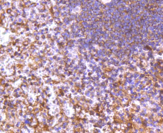
Immunohistochemical analysis of paraffin-embedded human spleen tissue using anti-DIABLO antibody. Counter stained with hematoxylin.IHC
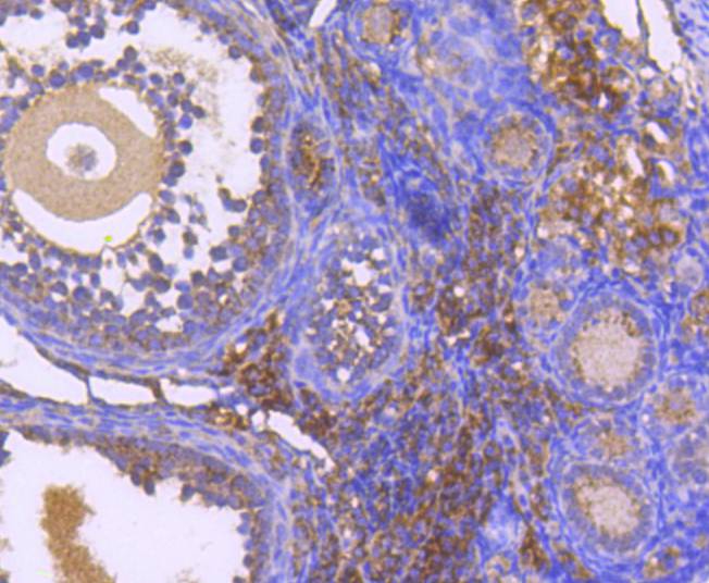
Immunohistochemical analysis of paraffin-embedded mouse overy tissue using anti-DIABLO antibody. Counter stained with hematoxylin.IHC
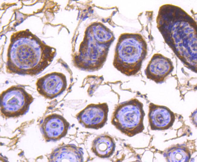
Immunohistochemical analysis of paraffin-embedded mouse skin tissue using anti-DIABLO antibody. Counter stained with hematoxylin.FC
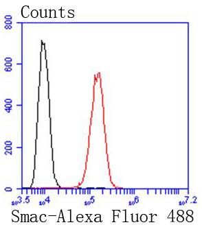
Flow cytometric analysis of HepG2 cells with DIABLO antibody at 1/50 dilution (red) compared with an unlabelled control (cells without incubation with primary antibody; black). Alexa Fluor 488-conjugated goat anti rabbit IgG was used as the secondary antibody.| Product Name | DIABLO Recombinant Rabbit Monoclonal Antibody |
|---|---|
| Antibody Type | Primary Antibodies |
| Product description | The activation of caspases is a key regulatory step in apoptosis. Once cytochrome c is released from the mitochondria into the cytosol, it binds Apaf-1 to form an oligomeric cytochrome c/Apaf-1 complex, which induces caspase activation. Inhibitors of Apoptosis Proteins (IAPs), are a family of proteins that regulate the cytochrome c/Apaf-1 caspase activating pathway. Like cytochrome c, Smac (for second mitochondria-derived activator of caspase, also designated DIABLO in mouse for direct IAP binding protein with low PI) promotes caspase activation in the cytochrome c/Apaf-1/caspase-9 pathway by binding IAPs and preventing them from inhibiting caspases. In healthy cells, Smac is a mitochondrial protein, but when cells undergo apoptosis, Smac is released into the cytosol. |
| Immunogen | Synthetic peptide within Human DIABLO aa 190-239 / 239. |
| Clonality | monoclonal |
|---|---|
| Isotype | IgG |
| Host Species | Recombinant rabbit |
| Tested Applications | FCICC/IFIHCWB |
| WB:1:2000 ICC/IF:1:50-1:200 IHC:1:50-1:200 FC:1:50-1:100 |
|
| Species Reactivity | HumanMouseRat |
| Concentration | 1mg/ml |
| Purification | Protein A |
| Alternative Names | 0610041G12Rik antibody DBLOH_HUMAN antibody DBOH antibody DFNA64 antibody diablo antibody Diablo homolog (Drosophila) antibody Diablo homolog antibody Diablo homolog mitochondrial antibody Diablo IAP binding mitochondrial protein antibody Diablo like protein antibody DIABLO S antibody Direct IAP binding protein with low pI antibody Direct IAP-binding protein with low pI antibody FLJ10537 antibody FLJ25049 antibody mitochondrial antibody Mitochondrial Smac protein antibody Second mitochondria derived activator of caspase antibody Second mitochondria-derived activator of caspase antibody SMAC 3 antibody Smac antibody Smac protein antibody SMAC3 antibody |
|---|---|
| Molecular Weight(MW) | 21kDa |
| Cellular Localization | Mitochondrion. |
WB
Western blot analysis of DIABLO on different lysates with Rabbit anti-DIABLO antibody at 1/2,000 dilution. Lane 1: HeLa cell lysate (20 µg/Lane), Lane 2: Jurkat cell lysate (20 µg/Lane), Lane 3: C6 cell lysate (20 µg/Lane), Lane 4: Mouse testis tissue lysate (40 µg/Lane), Lane 5: Rat testis tissue lysate (40 µg/Lane), Exposure time: 2 seconds; 4-20% SDS-PAGE gel. Proteins were transferred to a PVDF membrane and blocked with 5% NFDM/TBST for 1 hour at room temperature. The primary antibody at 1/2,000 dilution was used in 5% NFDM/TBST at 4℃ overnight. Goat Anti-Rabbit IgG - HRP Secondary Antibody at 1/50,000 dilution was used for 1 hour at room temperature.
ICC/IF
Immunocytochemistry analysis of HeLa cells labeling DIABLO with Rabbit anti-DIABLO antibody at 1/100 dilution. Cells were fixed in 4% paraformaldehyde for 15 minutes at room temperature, permeabilized with 0.1% Triton X-100 in PBS for 15 minutes at room temperature, then blocked with 1% BSA in 10% negative goat serum for 1 hour at room temperature. Cells were then incubated with Rabbit anti-DIABLO antibody at 1/100 dilution in 1% BSA in PBST overnight at 4 ℃. Goat Anti-Rabbit IgG H&L (488) was used as the secondary antibody at 1/1,000 dilution. PBS instead of the primary antibody was used as the secondary antibody only control. Counterstained with Mitotracker. Nuclear DNA was labelled in blue with DAPI.
ICC/IF
Immunocytochemistry analysis of Neuro-2a cells labeling DIABLO with Rabbit anti-DIABLO antibody at 1/100 dilution. Cells were fixed in 4% paraformaldehyde for 15 minutes at room temperature, permeabilized with 0.1% Triton X-100 in PBS for 15 minutes at room temperature, then blocked with 1% BSA in 10% negative goat serum for 1 hour at room temperature. Cells were then incubated with Rabbit anti-DIABLO antibody at 1/100 dilution in 1% BSA in PBST overnight at 4 ℃. Goat Anti-Rabbit IgG H&L (488) was used as the secondary antibody at 1/1,000 dilution. PBS instead of the primary antibody was used as the secondary antibody only control. Counterstained with Mitotracker. Nuclear DNA was labelled in blue with DAPI.
ICC/IF
Immunocytochemistry analysis of C6 cells labeling DIABLO with Rabbit anti-DIABLO antibody at 1/100 dilution. Cells were fixed in 4% paraformaldehyde for 15 minutes at room temperature, permeabilized with 0.1% Triton X-100 in PBS for 15 minutes at room temperature, then blocked with 1% BSA in 10% negative goat serum for 1 hour at room temperature. Cells were then incubated with Rabbit anti-DIABLO antibody at 1/100 dilution in 1% BSA in PBST overnight at 4 ℃. Goat Anti-Rabbit IgG H&L (488) was used as the secondary antibody at 1/1,000 dilution. PBS instead of the primary antibody was used as the secondary antibody only control. Counterstained with Mitotracker. Nuclear DNA was labelled in blue with DAPI.
IHC
Immunohistochemical analysis of paraffin-embedded human spleen tissue using anti-DIABLO antibody. Counter stained with hematoxylin.
IHC
Immunohistochemical analysis of paraffin-embedded mouse overy tissue using anti-DIABLO antibody. Counter stained with hematoxylin.
IHC
Immunohistochemical analysis of paraffin-embedded mouse skin tissue using anti-DIABLO antibody. Counter stained with hematoxylin.
FC
Flow cytometric analysis of HepG2 cells with DIABLO antibody at 1/50 dilution (red) compared with an unlabelled control (cells without incubation with primary antibody; black). Alexa Fluor 488-conjugated goat anti rabbit IgG was used as the secondary antibody.| Application Notes | WB:1:2000 ICC/IF:1:50-1:200 IHC:1:50-1:200 FC:1:50-1:100 |
|---|
| Form | Liquid |
|---|---|
| Storage Instructions | Store at +4℃ after thawing. Aliquot store at -20℃ or -80℃. Avoid repeated freeze / thaw cycles. |
| Storage Buffer | 1*TBS (pH7.4), 1%BSA, 40%Glycerol. Preservative: 0.05% Sodium Azide. |


