Application
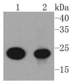
Fig1: Western blot analysis of AKAP14 on different lysates using anti-AKAP14 antibody at 1/1,000 dilution. Positive control: Lane 1: Jurkat Lane 2: A549Application
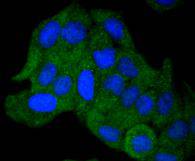
Fig2: ICC staining AKAP14 in Hela cells (green). The nuclear counter stain is DAPI (blue). Cells were fixed in paraformaldehyde, permeabilised with 0.25% Triton X100/PBS.Application
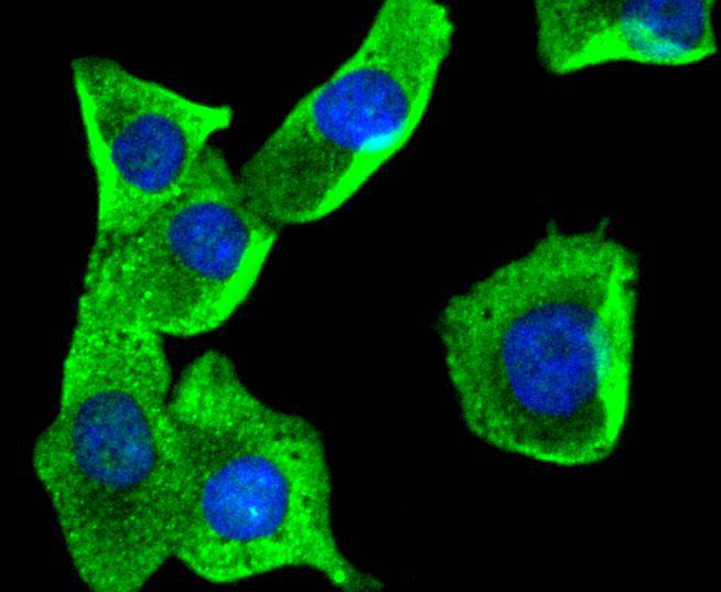
Fig3: ICC staining AKAP14 in A549 cells (green). The nuclear counter stain is DAPI (blue). Cells were fixed in paraformaldehyde, permeabilised with 0.25% Triton X100/PBS.Application
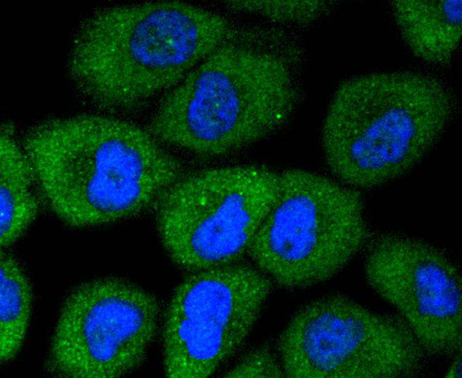
Fig4: ICC staining AKAP14 in HUVEC cells (green). The nuclear counter stain is DAPI (blue). Cells were fixed in paraformaldehyde, permeabilised with 0.25% Triton X100/PBS.| Product Name | AKAP14 [SC0667] |
|---|---|
| Antibody Type | Primary Antibodies |
| Antigen Alias | A kinase (PRKA) anchor protein 14 antibody A kinase anchor protein 14 antibody A kinase anchor protein 28 kDa antibody A-kinase anchoring protein 28 antibody AKAP14 antibody AKAP28 antibody PRKA14 antibody Protein kinase A anchoring protein 14 antibody |
| Product description | The A-kinase anchor proteins (AKAPs) are a group of structurally diverse proteins, which have the common function of binding to the regulatory subunit of protein kinase A (PKA) and confining the holoenzyme to discrete locations within the cell. This gene encodes a member of the AKAP family. The protein anchors PKA in ciliary axonemes and, in this way, may play a role in regulating ciliary beat frequency. Alternate transcriptional splice variants, encoding different isoforms, have been characterized. Binds to type II regulatory subunits of protein kinase A and anchors/targets them. |
| Immunogen | recombinant protein |
| Clonality | Monoclonal |
|---|---|
| Isotype | IgG |
| Host Species | Recombinant rabbit |
| Tested Applications | WBICC/IFIP |
| WB:1:1,000-1:5,000 ICC:1:50-1:200 |
|
| Species Reactivity | HumanMouseRat |
| Concentration | 1mg/ml |
| Alternative Names | A kinase (PRKA) anchor protein 14 antibody A kinase anchor protein 14 antibody A kinase anchor protein 28 kDa antibody A-kinase anchoring protein 28 antibody AKAP14 antibody AKAP28 antibody PRKA14 antibody Protein kinase A anchoring protein 14 antibody |
|---|---|
| Molecular Weight(MW) | 23 kDa |
| Cellular Localization | Cytoplasm. |
Application
Fig1: Western blot analysis of AKAP14 on different lysates using anti-AKAP14 antibody at 1/1,000 dilution. Positive control: Lane 1: Jurkat Lane 2: A549
Application
Fig2: ICC staining AKAP14 in Hela cells (green). The nuclear counter stain is DAPI (blue). Cells were fixed in paraformaldehyde, permeabilised with 0.25% Triton X100/PBS.
Application
Fig3: ICC staining AKAP14 in A549 cells (green). The nuclear counter stain is DAPI (blue). Cells were fixed in paraformaldehyde, permeabilised with 0.25% Triton X100/PBS.
Application
Fig4: ICC staining AKAP14 in HUVEC cells (green). The nuclear counter stain is DAPI (blue). Cells were fixed in paraformaldehyde, permeabilised with 0.25% Triton X100/PBS.| Positive Control | HUVEC, Jurkat, Hela, A549. |
|---|---|
| Application Notes | WB:1:1,000-1:5,000 ICC:1:50-1:200 |
| Form | Liquid |
|---|---|
| Storage Instructions | Store at +4℃ after thawing. Aliquot store at -20℃ or -80℃. Avoid repeated freeze / thaw cycles. |
| Storage Buffer | 1*TBS (pH7.4), 1%BSA, 40%Glycerol. Preservative: 0.05% Sodium Azide. |


