Application
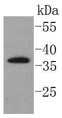
Fig1: Western blot analysis of SCF on Raji cells lysates using anti-SCF antibody at 1/1,000 dilution.Application
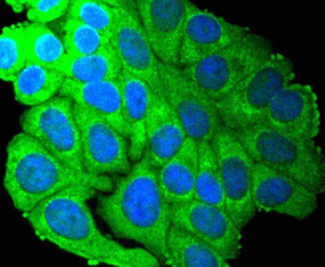
Fig2: ICC staining SCF in HepG2 cells (green). The nuclear counter stain is DAPI (blue). Cells were fixed in paraformaldehyde, permeabilised with 0.25% Triton X100/PBS.Application
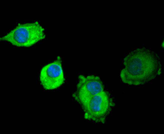
Fig3: ICC staining SCF in PANC-1 cells (green). The nuclear counter stain is DAPI (blue). Cells were fixed in paraformaldehyde, permeabilised with 0.25% Triton X100/PBS.Application
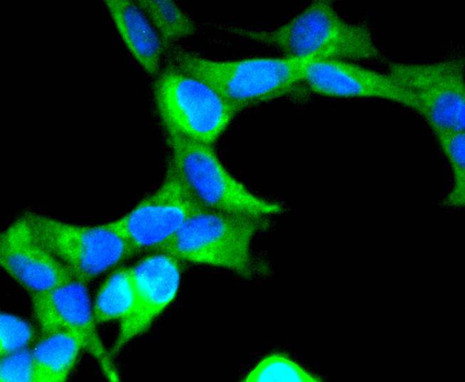
Fig4: ICC staining SCF in 293 cells (green). The nuclear counter stain is DAPI (blue). Cells were fixed in paraformaldehyde, permeabilised with 0.25% Triton X100/PBS.Application
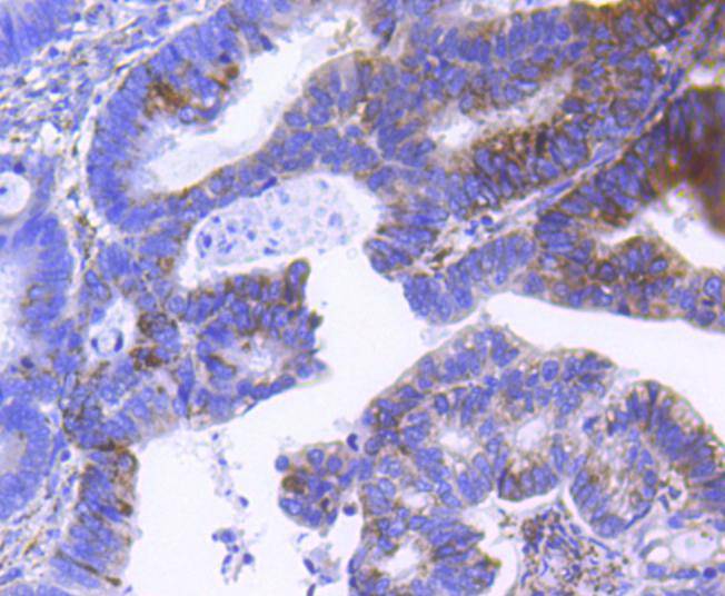
Fig5: Immunohistochemical analysis of paraffin-embedded human colon cancer tissue using anti-SCF antibody. Counter stained with hematoxylin.Application
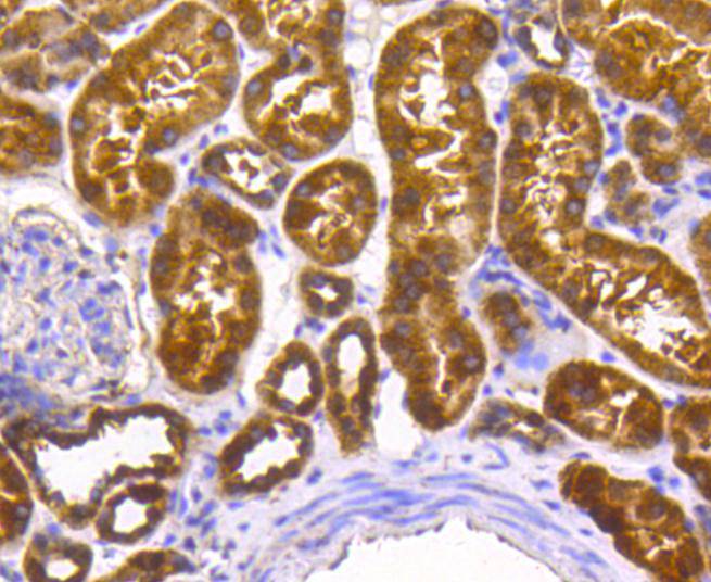
Fig6: Immunohistochemical analysis of paraffin-embedded human kidney tissue using anti-SCF antibody. Counter stained with hematoxylin.Application
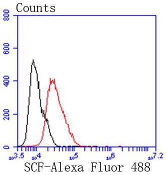
Fig7: Flow cytometric analysis of MCF-7 cells with SCF antibody at 1/50 dilution (red) compared with an unlabelled control (cells without incubation with primary antibody; black). Alexa Fluor 488-conjugated goat anti rabbit IgG was used as the secondary antibody.| Product Name | SCF [SC61-06] |
|---|---|
| Antibody Type | Primary Antibodies |
| Antigen Alias | C kit ligand antibody C-kit ligand antibody Ckit ligand antibody DCUA antibody DFNA69 antibody DKFZp686F2250 antibody familial progressive hyperpigmentation 2 antibody FPH2 antibody FPHH antibody KIT ligand antibody Kitl antibody KITLG antibody KL 1 antibody KL1 antibody Mast cell growth factor antibody MGF antibody MGF stem cell factor antibody SCF antibody SCF_HUMAN antibody SF antibody SHEP7 antibody sKITLG antibody Soluble KIT ligand antibody Steel factor antibody steel, mouse, homolog of antibody Stem cell factor antibody Stem cell factor precursor antibody |
| Product description | Stem cell factor (SCF) is the ligand for the transmembrane tyrosine kinase receptor proto-oncogene c-Kit. SCF, also designated KL, MGF and SLF, is a pleiotropic cytokine that has two alternatively spliced forms, 248 and 220 amino acids in length in human and mouse systems, respectively. Both the larger form and the smaller form are cleaved to produce soluble forms. The smaller splice variant lacks the proteolytic cleavage site, between amino acids 149 and 177 of the larger SCF form. The larger form of SCF is expressed in fibroblasts, brain and thymus, while the smaller form is found in spleen, testis, placenta and cerebellum. The SCF ligand is essential for the development of germ cells, hematopoietic progenitor cells and melanocyte precursors. SCF can stimulate the proliferation of mature mast cells as well as the maturation and proliferation of immature mast cells. |
| Immunogen | recombinant protein |
| Clonality | Monoclonal |
|---|---|
| Isotype | IgG |
| Host Species | Recombinant rabbit |
| Tested Applications | WBICC/IFIHCFC |
| WB:1:1,000-1:2,000 ICC:1:50-1:200 IHC:1:100-1:500 FC:1:50-1:100 |
|
| Species Reactivity | Human |
| Concentration | 1mg/ml |
| Alternative Names | C kit ligand antibody
C-kit ligand antibody
Ckit ligand antibody
DCUA antibody
DFNA69 antibody
DKFZp686F2250 antibody
familial progressive hyperpigmentation 2 antibody
FPH2 antibody
FPHH antibody
KIT ligand antibody
Kitl antibody
KITLG antibody
KL 1 antibody
KL1 antibody
Mast cell growth factor antibody
MGF antibody
MGF stem cell factor antibody
SCF antibody
SCF_HUMAN antibody
SF antibody
SHEP7 antibody
sKITLG antibody
Soluble KIT ligand antibody
Steel factor antibody
steel mouse homolog of antibody Stem cell factor antibody Stem cell factor precursor antibody |
|---|---|
| Molecular Weight(MW) | 37 kDa |
| Cellular Localization | Cytoplasm, Cell membrane, Secreted. |
| SwissProt ID | P21583 |
|---|
Application
Fig1: Western blot analysis of SCF on Raji cells lysates using anti-SCF antibody at 1/1,000 dilution.
Application
Fig2: ICC staining SCF in HepG2 cells (green). The nuclear counter stain is DAPI (blue). Cells were fixed in paraformaldehyde, permeabilised with 0.25% Triton X100/PBS.
Application
Fig3: ICC staining SCF in PANC-1 cells (green). The nuclear counter stain is DAPI (blue). Cells were fixed in paraformaldehyde, permeabilised with 0.25% Triton X100/PBS.
Application
Fig4: ICC staining SCF in 293 cells (green). The nuclear counter stain is DAPI (blue). Cells were fixed in paraformaldehyde, permeabilised with 0.25% Triton X100/PBS.
Application
Fig5: Immunohistochemical analysis of paraffin-embedded human colon cancer tissue using anti-SCF antibody. Counter stained with hematoxylin.
Application
Fig6: Immunohistochemical analysis of paraffin-embedded human kidney tissue using anti-SCF antibody. Counter stained with hematoxylin.
Application
Fig7: Flow cytometric analysis of MCF-7 cells with SCF antibody at 1/50 dilution (red) compared with an unlabelled control (cells without incubation with primary antibody; black). Alexa Fluor 488-conjugated goat anti rabbit IgG was used as the secondary antibody.| Positive Control | Raji, 293, PANC-1, HepG2, human kidney tissue, human colon cancer tissue. |
|---|---|
| Application Notes | WB:1:1,000-1:2,000 ICC:1:50-1:200 IHC:1:100-1:500 FC:1:50-1:100 |
| Form | Liquid |
|---|---|
| Storage Instructions | Store at +4℃ after thawing. Aliquot store at -20℃ or -80℃. Avoid repeated freeze / thaw cycles. |
| Storage Buffer | 1*TBS (pH7.4), 1%BSA, 40%Glycerol. Preservative: 0.05% Sodium Azide. |


