Application
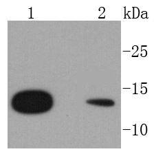
Fig1: Western blot analysis of RPS20 on different lysates using anti-RPS20 antibody at 1/1,000 dilution. Positive control: Lane 1: Hela Lane 2: A431Application
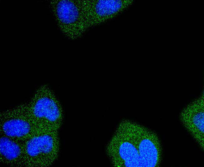
Fig2: ICC staining RPS20 in Hela cells (green). The nuclear counter stain is DAPI (blue). Cells were fixed in paraformaldehyde, permeabilised with 0.25% Triton X100/PBS.Application
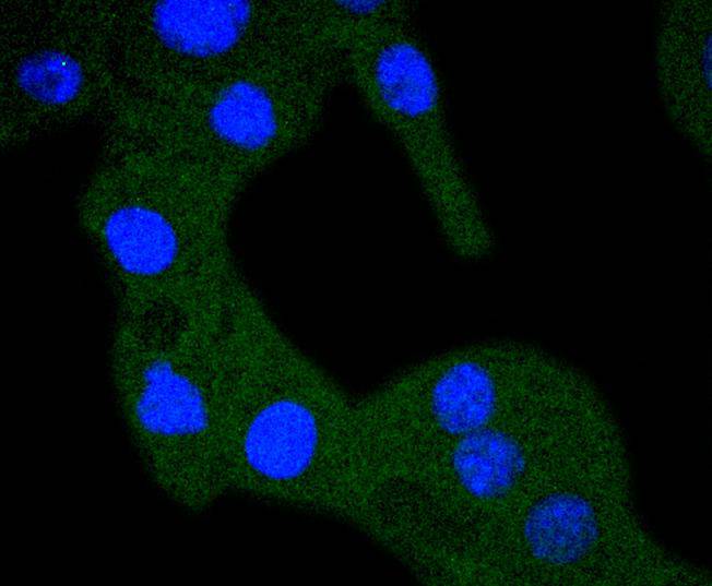
Fig3: ICC staining RPS20 in B16-F1 cells (green). The nuclear counter stain is DAPI (blue). Cells were fixed in paraformaldehyde, permeabilised with 0.25% Triton X100/PBS.| Product Name | RPS20 [SC65-08] |
|---|---|
| Antibody Type | Primary Antibodies |
| Antigen Alias | 40S ribosomal protein S20 antibody Ribosomal protein S20 antibody RPS 20 antibody RpS20 antibody RS20_HUMAN antibody S20 antibody |
| Product description | Mammalian ribosomal proteins are encoded by multigene families that consist of processed pseudogenes and one functional intron-containing gene within their coding regions. Ribosomal Protein S20, also known as RPS20, is a 119 amino acid cytoplasmic protein that is a component of the 40S ribosomal subunit. Co-transcribed with the small nucleolar RNA gene U54, Ribosomal Protein S20 is a primary binding protein (it binds independently to its target protein) that interacts with both the 5’ and 3’ minor domains of 16S ribosomal RNA (rRNA). Through its interactions with 16S rRNA, Ribosomal Protein S20 is thought to play a key role in nucleating the assembly of the 30S ribosomal subunit. Like most ribosomal protein-coding genes, the gene encoding Ribosomal Protein S20 is dispersed throughout the genome and exists as multiple processed pseudogenes. |
| Immunogen | recombinant protein |
| Clonality | Monoclonal |
|---|---|
| Isotype | IgG |
| Host Species | Recombinant rabbit |
| Tested Applications | WBICCIHC |
| WB:1:1,000-1:2,000 ICC:1:50-1:200 IHC:1:50-1:200 |
|
| Species Reactivity | HumanMouseRat |
| Concentration | 1mg/ml |
| Alternative Names | 40S ribosomal protein S20 antibody Ribosomal protein S20 antibody RPS 20 antibody RpS20 antibody RS20_HUMAN antibody S20 antibody |
|---|---|
| Molecular Weight(MW) | 13 kDa |
| Cellular Localization | Cytoplasm. |
Application
Fig1: Western blot analysis of RPS20 on different lysates using anti-RPS20 antibody at 1/1,000 dilution. Positive control: Lane 1: Hela Lane 2: A431
Application
Fig2: ICC staining RPS20 in Hela cells (green). The nuclear counter stain is DAPI (blue). Cells were fixed in paraformaldehyde, permeabilised with 0.25% Triton X100/PBS.
Application
Fig3: ICC staining RPS20 in B16-F1 cells (green). The nuclear counter stain is DAPI (blue). Cells were fixed in paraformaldehyde, permeabilised with 0.25% Triton X100/PBS.| Positive Control | A431, Hela, B16-F1. |
|---|---|
| Application Notes | WB:1:1,000-1:2,000 ICC:1:50-1:200 IHC:1:50-1:200 |
| Form | Liquid |
|---|---|
| Storage Instructions | Store at +4℃ after thawing. Aliquot store at -20℃ or -80℃. Avoid repeated freeze / thaw cycles. |
| Storage Buffer | 1*TBS (pH7.4), 1%BSA, 40%Glycerol. Preservative: 0.05% Sodium Azide. |


