Application
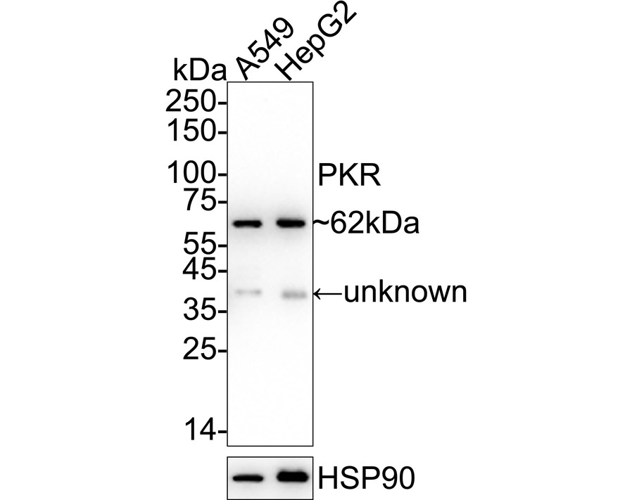
Fig1: Western blot analysis of PKR on different lysates with Rabbit anti-PKR antibody at 1/2,000 dilution. Lane 1: A549 cell lysate, Lane 2: HepG2 cell lysate, Lysates/proteins at 20 µg/Lane. Exposure time: 1 minute; 4-20% SDS-PAGE gel. Proteins were transferred to a PVDF membrane and blocked with 5% NFDM/TBST for 1 hour at room temperature. The primary antibody at 1/2,000 dilution was used in 5% NFDM/TBST at 4℃ overnight. Goat Anti-Rabbit IgG - HRP Secondary Antibody at 1/50,000 dilution was used for 1 hour at room temperature.Application
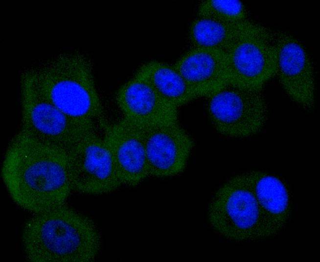
Fig2: ICC staining PKR in MCF-7 cells (green). The nuclear counter stain is DAPI (blue). Cells were fixed in paraformaldehyde, permeabilised with 0.25% Triton X100/PBS.Application
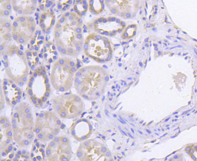
Fig3: Immunohistochemical analysis of paraffin-embedded human kidney tissue using anti-PKR antibody. Counter stained with hematoxylin.| Product Name | PKR Recombinant Rabbit Monoclonal Antibody |
|---|---|
| Antibody Type | Primary Antibodies |
| Product description | An interferon-inducible, RNA-dependent protein serine/threonine kinase (PKR) has been described. PKR in earlier literature is variously known as DAI, dsJ, PI kinase, p65, p67 or TIK for the mouse kinase; and p68 or p69 for the human kinase. The PKR kinase substrate is the a subunit of protein synthesis initiation factor eIF-2. Phosphorylation of eIF-2a on serine-51 results in inhibition of translation. Molecular cDNA clones have been isolated from both human and mouse cells. The serine/threonine kinase catalytic domains map to the carboxy terminal half of the protein while the RNA-binding domains are located in the amino terminal region. Three kinds of regulation of PKR enzymatic activity have been described. These include transcriptional regulation in response to interferon, an autoregulatory mechanism controlling PKR expression at the level of translation and post-translational regulation by RNA mediated autophosphorylation. |
| Immunogen | Synthetic peptide within human PKR aa 500-540. |
| Clonality | monoclonal |
|---|---|
| Isotype | IgG |
| Host Species | Recombinant rabbit |
| Tested Applications | ICC/IFIHCIPWB |
| WB:1:1,000-1:5,000 ICC:1:50-1:100 IHC:1:50-1:100 |
|
| Species Reactivity | Human |
| Concentration | 1mg/ml |
| Purification | Protein A |
| Alternative Names | Double stranded RNA activated protein kinase antibody E2AK2_HUMAN antibody eIF-2A protein kinase 2 antibody EIF2AK1 antibody EIF2AK2 antibody Eukaryotic translation initiation factor 2 alpha kinase 2 antibody Eukaryotic translation initiation factor 2-alpha kinase 2 antibody HGNC:9437 antibody Interferon induced double stranded RNA activated protein kinase antibody Interferon inducible elF2 alpha kinase antibody Interferon inducible RNA dependent protein kinase antibody Interferon-induced double-stranded RNA-activated protein kinase antibody Interferon-inducible RNA-dependent protein kinase antibody MGC126524 antibody P1/eIF-2A protein kinase antibody P1/eIF2A protein kinase antibody p68 kinase antibody PKR antibody PPP1R83 antibody PRKR antibody Protein kinase interferon inducible double stranded RNA dependent antibody Protein kinase RNA activated antibody Protein kinase RNA-activated antibody Protein phosphatase 1 regulatory subunit 83 antibody Serine/threonine protein kinase TIK antibody Tyrosine protein kinase EIF2AK2 antibody |
|---|---|
| Molecular Weight(MW) | 62kDa |
| Cellular Localization | Cytoplasm, Nucleus. |
| SwissProt ID | P19525 |
|---|
Application
Fig1: Western blot analysis of PKR on different lysates with Rabbit anti-PKR antibody at 1/2,000 dilution. Lane 1: A549 cell lysate, Lane 2: HepG2 cell lysate, Lysates/proteins at 20 µg/Lane. Exposure time: 1 minute; 4-20% SDS-PAGE gel. Proteins were transferred to a PVDF membrane and blocked with 5% NFDM/TBST for 1 hour at room temperature. The primary antibody at 1/2,000 dilution was used in 5% NFDM/TBST at 4℃ overnight. Goat Anti-Rabbit IgG - HRP Secondary Antibody at 1/50,000 dilution was used for 1 hour at room temperature.
Application
Fig2: ICC staining PKR in MCF-7 cells (green). The nuclear counter stain is DAPI (blue). Cells were fixed in paraformaldehyde, permeabilised with 0.25% Triton X100/PBS.
Application
Fig3: Immunohistochemical analysis of paraffin-embedded human kidney tissue using anti-PKR antibody. Counter stained with hematoxylin.| Positive Control | MCF-7, Hela, human kidney tissue. |
|---|---|
| Application Notes | WB:1:1,000-1:5,000 ICC:1:50-1:100 IHC:1:50-1:100 |
| Form | Liquid |
|---|---|
| Storage Instructions | Store at +4℃ after thawing. Aliquot store at -20℃ or -80℃. Avoid repeated freeze / thaw cycles. |
| Storage Buffer | 1*TBS (pH7.4), 1%BSA, 40%Glycerol. Preservative: 0.05% Sodium Azide. |


