WB
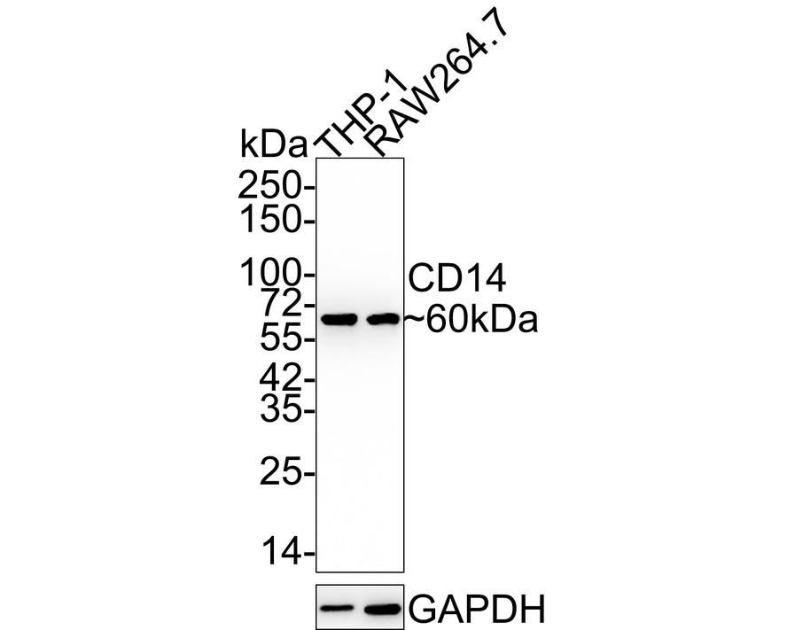
Western blot analysis of CD14 on different lysates with Rabbit anti-CD14 antibody at 1/5,000 dilution. Lane 1: THP-1 cell lysate, Lane 2: RAW264.7 cell lysate, Lysates/proteins at 15 µg/Lane. Exposure time: 1 minute 40 seconds; 4-20% SDS-PAGE gel. Proteins were transferred to a PVDF membrane and blocked with 5% NFDM/TBST for 1 hour at room temperature. The primary antibody at 1/5,000 dilution was used in 5% NFDM/TBST at 4℃ overnight. Goat Anti-Rabbit IgG - HRP Secondary Antibody at 1:50,000 dilution was used for 1 hour at room temperature.IHC
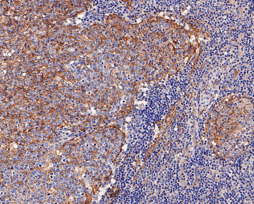
Immunohistochemical analysis of paraffin-embedded human lymph nodes tissue with Rabbit anti-CD14 antibody at 1/800 dilution. The section was pre-treated using heat mediated antigen retrieval with Tris-EDTA buffer (pH 9.0) for 20 minutes. The tissues were blocked in 1% BSA for 20 minutes at room temperature, washed with ddH2O and PBS, and then probed with the primary antibody at 1/800 dilution for 1 hour at room temperature. The detection was performed using an HRP conjugated compact polymer system. DAB was used as the chromogen. Tissues were counterstained with hematoxylin and mounted with DPX.IHC
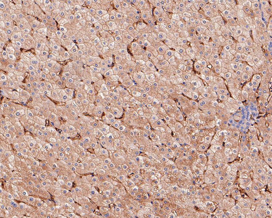
Immunohistochemical analysis of paraffin-embedded human liver tissue with Rabbit anti-CD14 antibody at 1/800 dilution. The section was pre-treated using heat mediated antigen retrieval with Tris-EDTA buffer (pH 9.0) for 20 minutes. The tissues were blocked in 1% BSA for 20 minutes at room temperature, washed with ddH2O and PBS, and then probed with the primary antibody at 1/800 dilution for 1 hour at room temperature. The detection was performed using an HRP conjugated compact polymer system. DAB was used as the chromogen. Tissues were counterstained with hematoxylin and mounted with DPX.ICC/IF
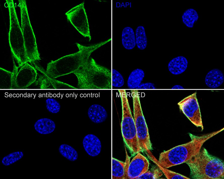
Immunocytochemistry analysis of NIH/3T3 cells labeling CD14 with Rabbit anti-CD14 antibody at 1/100 dilution. Cells were fixed in 4% paraformaldehyde for 20 minutes at room temperature, permeabilized with 0.1% Triton X-100 in PBS for 5 minutes at room temperature, then blocked with 1% BSA in 10% negative goat serum for 1 hour at room temperature. Cells were then incubated with Rabbit anti-CD14 antibody at 1/100 dilution in 1% BSA in PBST overnight at 4 ℃. Goat Anti-Rabbit IgG H&L (488) was used as the secondary antibody at 1/1,000 dilution. PBS instead of the primary antibody was used as the secondary antibody only control. Nuclear DNA was labelled in blue with DAPI. Beta tubulin ( red) was stained at 1/100 dilution overnight at +4℃. Goat Anti-Mouse IgG H&L (594) was used as the secondary antibody at 1/1,000 dilution.mIHC
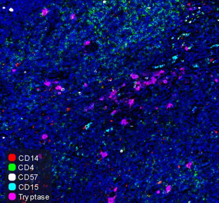
Fluorescence multiplex immunohistochemical analysis of Human tonsil (Formalin/PFA-fixed paraffin-embedded sections). Panel A: the merged image of anti-CD14 (Red), anti-CD4 (Green), anti-CD57 (White), anti-CD15 (Cyan)and anti-Tryptase (Magenta) on tonsil. Panel B: anti- CD14 stained on monocytes. Panel C: anti-CD4 stained on helper T cells and Treg cells. Panel D: anti-CD57 stained on NK cells and T cells. Panel E: CD15 stained on granulocytes and monocytes. Panel F: anti-Tryptase stained on Mast cells. HRP Conjugated UltraPolymer Goat Polyclonal Antibody was used as a secondary antibody. The immunostaining was performed with the Sequential Immuno-staining Kit (IRISKit™MH010101, www.luminiris.cn). The section was incubated in five rounds of staining: in the order of anti-CD14 (1/800 dilution), anti-CD4 (1/800 dilution), anti-CD57 (1/1,000 dilution), anti-CD15 (1/500 dilution), and anti-Tryptase (1/3,000 dilution) for 20 mins at room temperature. Each round was followed by a separate fluorescent tyramide signal amplification system. Heat mediated antigen retrieval with Tris-EDTA buffer (pH 9.0) for 30 mins at 95℃. DAPI (blue) was used as a nuclear counter stain. Image acquisition was performed with Olympus VS200 Slide Scanner.FC
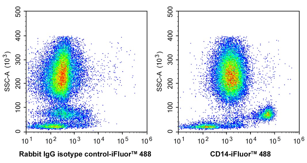
Flow cytometric analysis of human peripheral blood cells labelling CD14.| Product Name | CD14 Recombinant Rabbit Monoclonal Antibody |
|---|---|
| Antibody Type | Primary Antibodies |
| Product description | Lipopolysaccharide (LPS) elicits the secretion of mediators and cytokines produced by activated macrophages and monocytes. CD14 is a glycosylphosphatidylinositol (GPI)-anchored protein found on the surfaces of monocytes and polymorphonuclear leukocytes. CD14 functions as a receptor for LPS, resulting in the secretion of various proteins. An important component in the LPS activation of monocytes through the CD14 receptor is the "adapter molecule," lipopolysaccharide binding protein (LBP). There are two forms of CD14, a membrane-associated form (mCD14), and a soluble form (sCD14). mCD14 responds to LPS alone and facilitates the secretion of proteins, while cells not expressing mCD14 fail to respond to LPS. The cells that lack mCD14 respond to LPS/LBP in the presence of sCD14. |
| Immunogen | Synthetic peptide within Human CD14 aa 310-335 / 375. |
| Clonality | Monoclonal |
|---|---|
| Isotype | IgG |
| Host Species | Recombinant rabbit |
| Tested Applications | FCICC/IFIHCWBmIHC |
| WB:1:5000 IHC:1:200-1:800 ICC/IF:1:50-1:200 mIHC:1:800-1:1000 FC:1:1000 |
|
| Species Reactivity | HumanMouse |
| Concentration | 1mg/ml |
| Purification | Protein A |
| Alternative Names | CD 14 antibody
CD_antigen=CD14 antibody
CD14 antibody
CD14 antigen antibody
CD14 molecule antibody
CD14_HUMAN antibody
LPS-R antibody
Mo2 antibody
Monocyte differentiation antigen CD14 antibody
Monocyte differentiation antigen CD14 urinary form antibody
Monocyte differentiation antigen CD14 membrane-bound form antibody Myeloid cell specific leucine rich glycoprotein antibody Myeloid cell-specific leucine-rich glycoprotein antibody |
|---|---|
| Molecular Weight(MW) | 40 kDa(Observed band size: 60 kDa) |
| Cellular Localization | Cell membrane, Secreted, Membrane raft, Golgi apparatus. |
| SwissProt ID | P08571 |
|---|
WB
Western blot analysis of CD14 on different lysates with Rabbit anti-CD14 antibody at 1/5,000 dilution. Lane 1: THP-1 cell lysate, Lane 2: RAW264.7 cell lysate, Lysates/proteins at 15 µg/Lane. Exposure time: 1 minute 40 seconds; 4-20% SDS-PAGE gel. Proteins were transferred to a PVDF membrane and blocked with 5% NFDM/TBST for 1 hour at room temperature. The primary antibody at 1/5,000 dilution was used in 5% NFDM/TBST at 4℃ overnight. Goat Anti-Rabbit IgG - HRP Secondary Antibody at 1:50,000 dilution was used for 1 hour at room temperature.
IHC
Immunohistochemical analysis of paraffin-embedded human lymph nodes tissue with Rabbit anti-CD14 antibody at 1/800 dilution. The section was pre-treated using heat mediated antigen retrieval with Tris-EDTA buffer (pH 9.0) for 20 minutes. The tissues were blocked in 1% BSA for 20 minutes at room temperature, washed with ddH2O and PBS, and then probed with the primary antibody at 1/800 dilution for 1 hour at room temperature. The detection was performed using an HRP conjugated compact polymer system. DAB was used as the chromogen. Tissues were counterstained with hematoxylin and mounted with DPX.
IHC
Immunohistochemical analysis of paraffin-embedded human liver tissue with Rabbit anti-CD14 antibody at 1/800 dilution. The section was pre-treated using heat mediated antigen retrieval with Tris-EDTA buffer (pH 9.0) for 20 minutes. The tissues were blocked in 1% BSA for 20 minutes at room temperature, washed with ddH2O and PBS, and then probed with the primary antibody at 1/800 dilution for 1 hour at room temperature. The detection was performed using an HRP conjugated compact polymer system. DAB was used as the chromogen. Tissues were counterstained with hematoxylin and mounted with DPX.
ICC/IF
Immunocytochemistry analysis of NIH/3T3 cells labeling CD14 with Rabbit anti-CD14 antibody at 1/100 dilution. Cells were fixed in 4% paraformaldehyde for 20 minutes at room temperature, permeabilized with 0.1% Triton X-100 in PBS for 5 minutes at room temperature, then blocked with 1% BSA in 10% negative goat serum for 1 hour at room temperature. Cells were then incubated with Rabbit anti-CD14 antibody at 1/100 dilution in 1% BSA in PBST overnight at 4 ℃. Goat Anti-Rabbit IgG H&L (488) was used as the secondary antibody at 1/1,000 dilution. PBS instead of the primary antibody was used as the secondary antibody only control. Nuclear DNA was labelled in blue with DAPI. Beta tubulin ( red) was stained at 1/100 dilution overnight at +4℃. Goat Anti-Mouse IgG H&L (594) was used as the secondary antibody at 1/1,000 dilution.
mIHC
Fluorescence multiplex immunohistochemical analysis of Human tonsil (Formalin/PFA-fixed paraffin-embedded sections). Panel A: the merged image of anti-CD14 (Red), anti-CD4 (Green), anti-CD57 (White), anti-CD15 (Cyan)and anti-Tryptase (Magenta) on tonsil. Panel B: anti- CD14 stained on monocytes. Panel C: anti-CD4 stained on helper T cells and Treg cells. Panel D: anti-CD57 stained on NK cells and T cells. Panel E: CD15 stained on granulocytes and monocytes. Panel F: anti-Tryptase stained on Mast cells. HRP Conjugated UltraPolymer Goat Polyclonal Antibody was used as a secondary antibody. The immunostaining was performed with the Sequential Immuno-staining Kit (IRISKit™MH010101, www.luminiris.cn). The section was incubated in five rounds of staining: in the order of anti-CD14 (1/800 dilution), anti-CD4 (1/800 dilution), anti-CD57 (1/1,000 dilution), anti-CD15 (1/500 dilution), and anti-Tryptase (1/3,000 dilution) for 20 mins at room temperature. Each round was followed by a separate fluorescent tyramide signal amplification system. Heat mediated antigen retrieval with Tris-EDTA buffer (pH 9.0) for 30 mins at 95℃. DAPI (blue) was used as a nuclear counter stain. Image acquisition was performed with Olympus VS200 Slide Scanner.
FC
Flow cytometric analysis of human peripheral blood cells labelling CD14.| Application Notes | WB:1:5000 IHC:1:200-1:800 ICC/IF:1:50-1:200 mIHC:1:800-1:1000 FC:1:1000 |
|---|
| Form | Liquid |
|---|---|
| Storage Instructions | Store at +4℃ after thawing. Aliquot store at -20℃ or -80℃. Avoid repeated freeze / thaw cycles. |
| Storage Buffer | 1*TBS (pH7.4), 1%BSA, 40%Glycerol. Preservative: 0.05% Sodium Azide. |


