Application
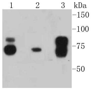
Fig1: Western blot analysis of Cortactin on different lysates using anti-Cortactin antibody at 1/1,000 dilution. Positive control: Lane 1: Hela Lane 2: NIH/3T3 Lane 3: A431Application
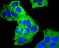
Fig2: ICC staining Cortactin in Hela cells (green). The nuclear counter stain is DAPI (blue). Cells were fixed in paraformaldehyde, permeabilised with 0.25% Triton X100/PBS.Application
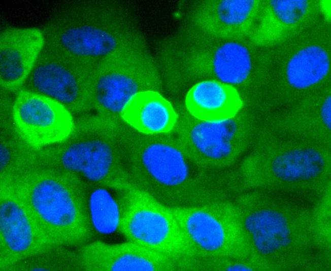
Fig3: ICC staining Cortactin in A431 cells (green). The nuclear counter stain is DAPI (blue). Cells were fixed in paraformaldehyde, permeabilised with 0.25% Triton X100/PBS.Application
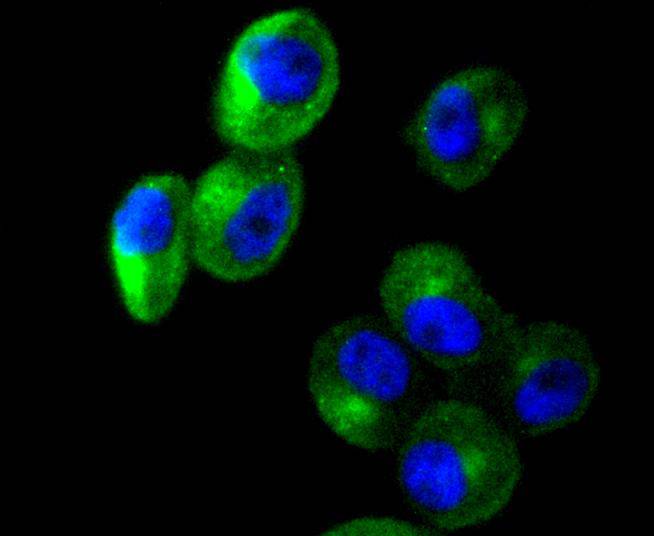
Fig4: ICC staining Cortactin in PC-3M cells (green). The nuclear counter stain is DAPI (blue). Cells were fixed in paraformaldehyde, permeabilised with 0.25% Triton X100/PBS.Application
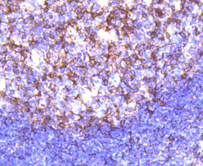
Fig5: Immunohistochemical analysis of paraffin-embedded human tonsil tissue using anti-Cortactin antibody. Counter stained with hematoxylin.Application
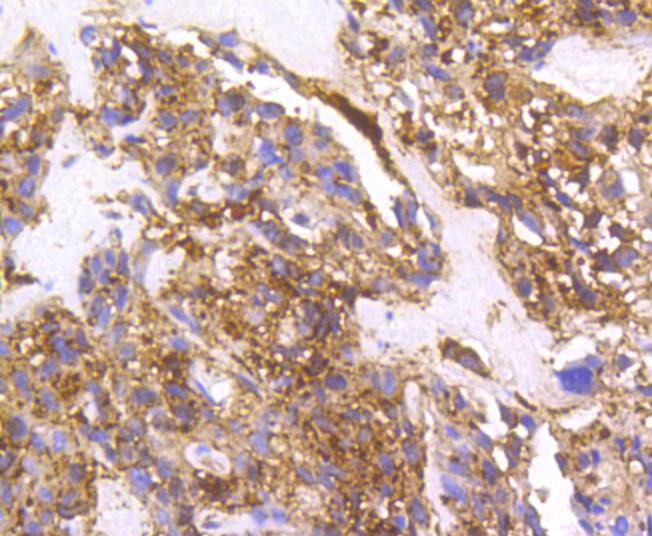
Fig6: Immunohistochemical analysis of paraffin-embedded human breast carcinoma tissue using anti-Cortactin antibody. Counter stained with hematoxylin.Application
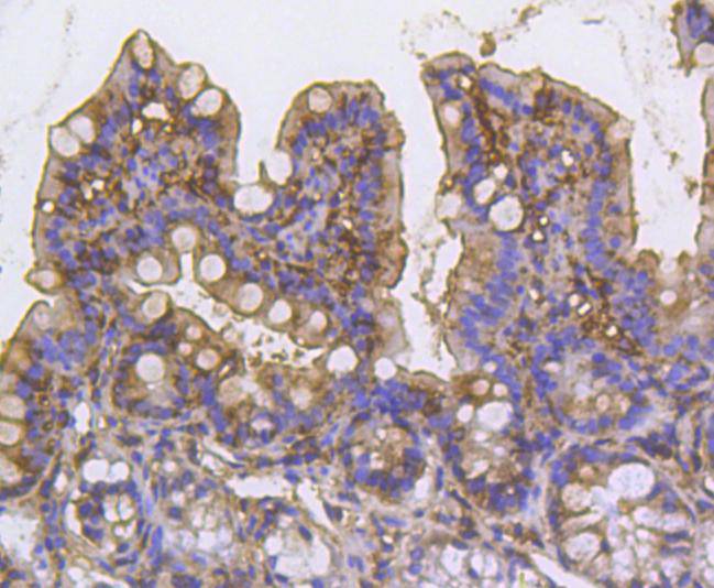
Fig7: Immunohistochemical analysis of paraffin-embedded mouse colon tissue using anti-Cortactin antibody. Counter stained with hematoxylin.Application
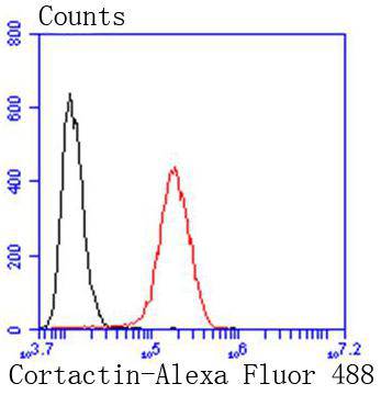
Fig8: Flow cytometric analysis of Hela cells with Cortactin antibody at 1/50 dilution (red) compared with an unlabelled control (cells without incubation with primary antibody; black). Alexa Fluor 488-conjugated goat anti rabbit IgG was used as the secondary antibody.| Product Name | Cortactin [SC61-08] |
|---|---|
| Antibody Type | Primary Antibodies |
| Antigen Alias | Amplaxin antibody CTTN antibody EMS 1 antibody EMS1 antibody FLJ34459 antibody Mammary tumor and squamous cell carcinoma associated antibody Oncogene EMS1 antibody p80/85 src substrate antibody Src substrate cortactin antibody SRC8_HUMAN antibody |
| Product description | Cortactin (also designated Ems-1) is a filamentous Actin (F-Actin) binding protein that has been shown to be a substrate for Src p60. Cortactin contains tandem 37 amino acid repeats at the amino-terminus and an SH3 domain at the carboxy-terminus. The tandem repeats appear to be necessary for F-Actin binding. Tyrosine phosphorylation of Cortactin by Src p60 results in diminished F-Actin binding to Cortactin and reduced F-Actin cross-linking activity. Cortactin has also been shown to be phosphorylated in response to FGF-1. Cortactin exhibits abundant expression in megakaryocytes and platelets, and it may play a role in the maturation of megakaryocytes. |
| Immunogen | recombinant protein |
| Clonality | Monoclonal |
|---|---|
| Isotype | IgG |
| Host Species | Recombinant rabbit |
| Tested Applications | WBICC/IFIHCIPFC |
| WB:1:1,000-1:5,000 ICC:1:100-1:500 IHC:1:50-1:200 FC:1:50-1:100 |
|
| Species Reactivity | HumanMouseRat |
| Concentration | 1mg/ml |
| Alternative Names | Amplaxin antibody CTTN antibody EMS 1 antibody EMS1 antibody FLJ34459 antibody Mammary tumor and squamous cell carcinoma associated antibody Oncogene EMS1 antibody p80/85 src substrate antibody Src substrate cortactin antibody SRC8_HUMAN antibody |
|---|---|
| Molecular Weight(MW) | 75 kDa |
| Cellular Localization | Cytoplasm, Cell projection, Membrane. |
Application
Fig1: Western blot analysis of Cortactin on different lysates using anti-Cortactin antibody at 1/1,000 dilution. Positive control: Lane 1: Hela Lane 2: NIH/3T3 Lane 3: A431
Application
Fig2: ICC staining Cortactin in Hela cells (green). The nuclear counter stain is DAPI (blue). Cells were fixed in paraformaldehyde, permeabilised with 0.25% Triton X100/PBS.
Application
Fig3: ICC staining Cortactin in A431 cells (green). The nuclear counter stain is DAPI (blue). Cells were fixed in paraformaldehyde, permeabilised with 0.25% Triton X100/PBS.
Application
Fig4: ICC staining Cortactin in PC-3M cells (green). The nuclear counter stain is DAPI (blue). Cells were fixed in paraformaldehyde, permeabilised with 0.25% Triton X100/PBS.
Application
Fig5: Immunohistochemical analysis of paraffin-embedded human tonsil tissue using anti-Cortactin antibody. Counter stained with hematoxylin.
Application
Fig6: Immunohistochemical analysis of paraffin-embedded human breast carcinoma tissue using anti-Cortactin antibody. Counter stained with hematoxylin.
Application
Fig7: Immunohistochemical analysis of paraffin-embedded mouse colon tissue using anti-Cortactin antibody. Counter stained with hematoxylin.
Application
Fig8: Flow cytometric analysis of Hela cells with Cortactin antibody at 1/50 dilution (red) compared with an unlabelled control (cells without incubation with primary antibody; black). Alexa Fluor 488-conjugated goat anti rabbit IgG was used as the secondary antibody.| Positive Control | NIH/3T3, A431, Hela, PC-3M, mouse colon tissue, human tonsil tissue, human breast carcinoma tissue. |
|---|---|
| Application Notes | WB:1:1,000-1:5,000 ICC:1:100-1:500 IHC:1:50-1:200 FC:1:50-1:100 |
| Form | Liquid |
|---|---|
| Storage Instructions | Store at +4℃ after thawing. Aliquot store at -20℃ or -80℃. Avoid repeated freeze / thaw cycles. |
| Storage Buffer | 1*TBS (pH7.4), 1%BSA, 40%Glycerol. Preservative: 0.05% Sodium Azide. |


