WB
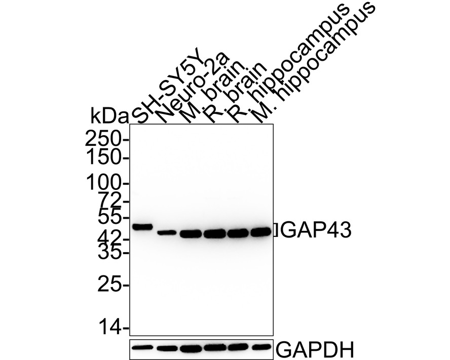
Western blot analysis of GAP43 on different lysates with Rabbit anti-GAP43 antibody at 1/20,000 dilution. Lane 1: SH-SY5Y cell lysate, Lane 2: Neuro-2a cell lysate, Lane 3: Mouse brain tissue lysate, Lane 4: Rat brain tissue lysate, Lane 5: Rat hippocampus tissue lysate, Lane 6: Mouse hippocampus tissue lysate, Lysates/proteins at 20 µg/Lane. Exposure time: 24 seconds; 4-20% SDS-PAGE gel. Proteins were transferred to a PVDF membrane and blocked with 5% NFDM/TBST for 1 hour at room temperature. The primary antibody at 1/20,000 dilution was used in 5% NFDM/TBST at 4℃ overnight. Goat Anti-Rabbit IgG - HRP Secondary Antibody at 1:50,000 dilution was used for 1 hour at room temperature.IHC
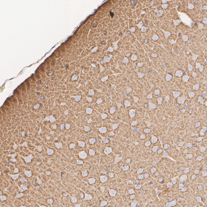
Immunohistochemical analysis of paraffin-embedded mouse brain tissue with Rabbit anti-GAP43 antibody at 1/15,000 dilution. The section was pre-treated using heat mediated antigen retrieval with Tris-EDTA buffer (pH 9.0) for 20 minutes. The tissues were blocked in 1% BSA for 20 minutes at room temperature, washed with ddH2O and PBS, and then probed with the primary antibody at 1/15,000 dilution for 1 hour at room temperature. The detection was performed using an HRP conjugated compact polymer system. DAB was used as the chromogen. Tissues were counterstained with hematoxylin and mounted with DPX.IHC
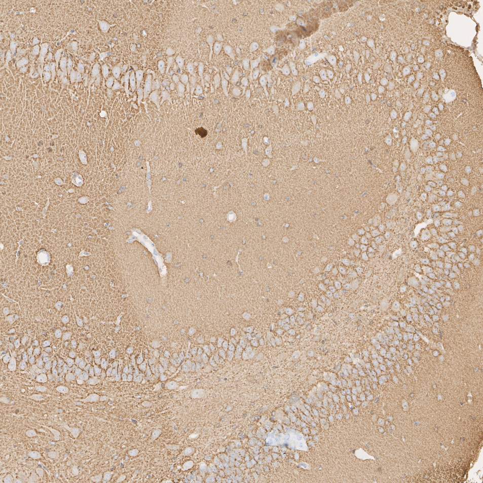
Immunohistochemical analysis of paraffin-embedded rat hippocampus tissue with Rabbit anti-GAP43 antibody at 1/15,000 dilution. The section was pre-treated using heat mediated antigen retrieval with Tris-EDTA buffer (pH 9.0) for 20 minutes. The tissues were blocked in 1% BSA for 20 minutes at room temperature, washed with ddH2O and PBS, and then probed with the primary antibody at 1/15,000 dilution for 1 hour at room temperature. The detection was performed using an HRP conjugated compact polymer system. DAB was used as the chromogen. Tissues were counterstained with hematoxylin and mounted with DPX.ICC/IF
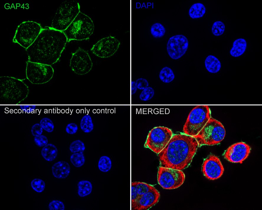
Immunocytochemistry analysis of Neuro-2a cells labeling GAP43 with Rabbit anti-GAP43 antibody at 1/500 dilution. Cells were fixed in 100% precooled methanol for 5 minutes at room temperature, then blocked with 1% BSA in 10% negative goat serum for 1 hour at room temperature. Cells were then incubated with Rabbit anti-GAP43 antibody at 1/500 dilution in 1% BSA in PBST overnight at 4 ℃. Goat Anti-Rabbit IgG H&L (488) was used as the secondary antibody at 1/1,000 dilution. PBS instead of the primary antibody was used as the secondary antibody only control. Nuclear DNA was labelled in blue with DAPI. Beta tubulin (red) was stained at 1/100 dilution overnight at +4℃. Goat Anti-Mouse IgG H&L (594) was used as the secondary antibody at 1/1,000 dilution.IF-P
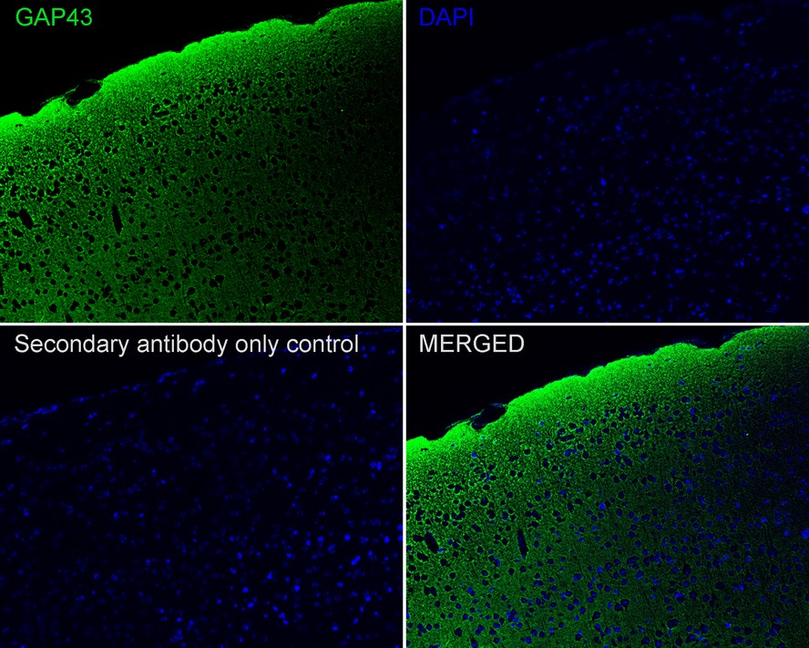
Immunofluorescence analysis of paraffin-embedded mouse brain tissue labeling GAP43 with Rabbit anti-GAP43 antibody at 1/200 dilution. The section was pre-treated using heat mediated antigen retrieval with Tris-EDTA buffer (pH 9.0) for 20 minutes. The tissues were blocked in 10% negative goat serum for 1 hour at room temperature, washed with PBS, and then probed with the primary antibody (green) at 1/200 dilution overnight at 4 ℃, washed with PBS. Goat Anti-Rabbit IgG H&L (488) was used as the secondary antibody at 1/1,000 dilution. Nuclei were counterstained with DAPI (blue).FC
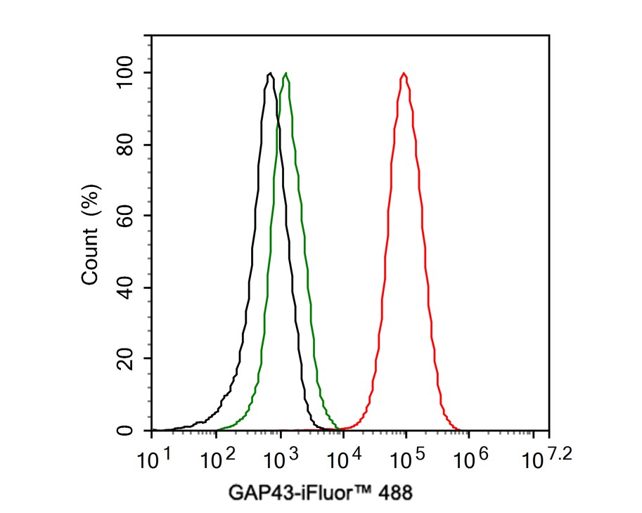
Intracellular Flow Cytometry analysis of SH-SY5Y labeling GAP43 with purified at 1/1,000 dilution (1 µg/ml) (red). Cells were fixed with 4% PFA and permeabilised with 90% methanol. Rabbit monoclonal IgG (green) was used as the isotype control, cells without incubation with primary antibody and secondary antibody (black) were used as the unlabeled control. A Goat anti-rabbit IgG 488 (1/1,000 dilution) was used as the secondary antibody.| Product Name | GAP43 Recombinant Rabbit Monoclonal Antibody |
|---|---|
| Antibody Type | Primary Antibodies |
| Product description | GAP-43 (growth associated protein 43, B-50, PP46, calmodulin-binding protein P-57, neuromodulin, neuron growth-associated protein 43, protein F1 is a crucial component for regenerative response in the nervous system. It is present at high levels in neuronal growth cones during development and axonal regeneration. GAP-43 is normally produced by neurons during developmental growth and axonal regeneration, but it is also expressed in specific regions of the normal adult nervous system. The neuron-specific Elav/Hu family member, HuD, interacts with and stabilizes GAP-43 mRNA in developing neurons and leads to increased levels of GAP-43 protein. Heterozygous GAP-43 knockout mice with GAP-43 levels reduced by one-half dipslay significant memory impairments in cued conditioning or on tests of nociceptive or auditory perception. |
| Immunogen | Synthetic peptide within Human GAP43 aa 189-238 / 238. |
| Clonality | Monoclonal |
|---|---|
| Isotype | IgG |
| Host Species | Recombinant rabbit |
| Tested Applications | FCICC/IFIF-PIHCWB |
| WB:1:20000-1:100000 IHC:1:10000-1:15000 ICC/IF:1:500 IF-P:1:500-1:1000 FC:1:1000 |
|
| Species Reactivity | HumanMouseRat |
| Concentration | 1mg/ml |
| Purification | Protein A |
| Alternative Names | Axonal membrane protein GAP 43 antibody Axonal membrane protein GAP-43 antibody B 50 antibody Calmodulin binding protein P 57 antibody F1 antibody GAP 43 antibody GAP43 antibody Growth Associated Protein 43 antibody Growth-associated protein 43 antibody Nerve Growth Related Peptide antibody Nerve growth related peptide GAP43 antibody NEUM_HUMAN antibody Neural phosphoprotein B 50 antibody Neural phosphoprotein B-50 antibody Neuromodulin antibody Neuron growth associated protein 43 antibody PP46 antibody Protein F1 antibody QtrA-11580 antibody QtrA-13071 antibody |
|---|---|
| Molecular Weight(MW) | 25 kDa(Observed band size: 43/45 kDa) |
| Cellular Localization | Cell membrane, growth cone membrane, filopodium membrane, Cytoplasm, synapse, perikaryon, dendrite, axon. |
WB
Western blot analysis of GAP43 on different lysates with Rabbit anti-GAP43 antibody at 1/20,000 dilution. Lane 1: SH-SY5Y cell lysate, Lane 2: Neuro-2a cell lysate, Lane 3: Mouse brain tissue lysate, Lane 4: Rat brain tissue lysate, Lane 5: Rat hippocampus tissue lysate, Lane 6: Mouse hippocampus tissue lysate, Lysates/proteins at 20 µg/Lane. Exposure time: 24 seconds; 4-20% SDS-PAGE gel. Proteins were transferred to a PVDF membrane and blocked with 5% NFDM/TBST for 1 hour at room temperature. The primary antibody at 1/20,000 dilution was used in 5% NFDM/TBST at 4℃ overnight. Goat Anti-Rabbit IgG - HRP Secondary Antibody at 1:50,000 dilution was used for 1 hour at room temperature.
IHC
Immunohistochemical analysis of paraffin-embedded mouse brain tissue with Rabbit anti-GAP43 antibody at 1/15,000 dilution. The section was pre-treated using heat mediated antigen retrieval with Tris-EDTA buffer (pH 9.0) for 20 minutes. The tissues were blocked in 1% BSA for 20 minutes at room temperature, washed with ddH2O and PBS, and then probed with the primary antibody at 1/15,000 dilution for 1 hour at room temperature. The detection was performed using an HRP conjugated compact polymer system. DAB was used as the chromogen. Tissues were counterstained with hematoxylin and mounted with DPX.
IHC
Immunohistochemical analysis of paraffin-embedded rat hippocampus tissue with Rabbit anti-GAP43 antibody at 1/15,000 dilution. The section was pre-treated using heat mediated antigen retrieval with Tris-EDTA buffer (pH 9.0) for 20 minutes. The tissues were blocked in 1% BSA for 20 minutes at room temperature, washed with ddH2O and PBS, and then probed with the primary antibody at 1/15,000 dilution for 1 hour at room temperature. The detection was performed using an HRP conjugated compact polymer system. DAB was used as the chromogen. Tissues were counterstained with hematoxylin and mounted with DPX.
ICC/IF
Immunocytochemistry analysis of Neuro-2a cells labeling GAP43 with Rabbit anti-GAP43 antibody at 1/500 dilution. Cells were fixed in 100% precooled methanol for 5 minutes at room temperature, then blocked with 1% BSA in 10% negative goat serum for 1 hour at room temperature. Cells were then incubated with Rabbit anti-GAP43 antibody at 1/500 dilution in 1% BSA in PBST overnight at 4 ℃. Goat Anti-Rabbit IgG H&L (488) was used as the secondary antibody at 1/1,000 dilution. PBS instead of the primary antibody was used as the secondary antibody only control. Nuclear DNA was labelled in blue with DAPI. Beta tubulin (red) was stained at 1/100 dilution overnight at +4℃. Goat Anti-Mouse IgG H&L (594) was used as the secondary antibody at 1/1,000 dilution.
IF-P
Immunofluorescence analysis of paraffin-embedded mouse brain tissue labeling GAP43 with Rabbit anti-GAP43 antibody at 1/200 dilution. The section was pre-treated using heat mediated antigen retrieval with Tris-EDTA buffer (pH 9.0) for 20 minutes. The tissues were blocked in 10% negative goat serum for 1 hour at room temperature, washed with PBS, and then probed with the primary antibody (green) at 1/200 dilution overnight at 4 ℃, washed with PBS. Goat Anti-Rabbit IgG H&L (488) was used as the secondary antibody at 1/1,000 dilution. Nuclei were counterstained with DAPI (blue).
FC
Intracellular Flow Cytometry analysis of SH-SY5Y labeling GAP43 with purified at 1/1,000 dilution (1 µg/ml) (red). Cells were fixed with 4% PFA and permeabilised with 90% methanol. Rabbit monoclonal IgG (green) was used as the isotype control, cells without incubation with primary antibody and secondary antibody (black) were used as the unlabeled control. A Goat anti-rabbit IgG 488 (1/1,000 dilution) was used as the secondary antibody.| Application Notes | WB:1:20000-1:100000 IHC:1:10000-1:15000 ICC/IF:1:500 IF-P:1:500-1:1000 FC:1:1000 |
|---|
| Form | Liquid |
|---|---|
| Storage Instructions | Store at +4℃ after thawing. Aliquot store at -20℃ or -80℃. Avoid repeated freeze / thaw cycles. |
| Storage Buffer | 1*TBS (pH7.4), 1%BSA, 40%Glycerol. Preservative: 0.05% Sodium Azide. |


