WB
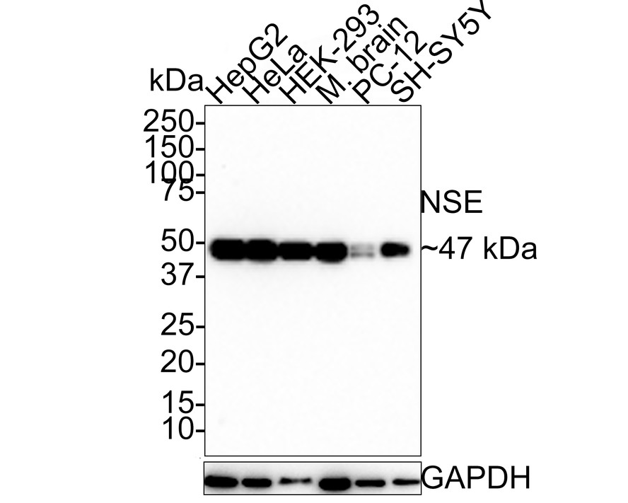
Western blot analysis of NSE on different lysates with Rabbit anti-NSE antibody at 1/1,000 dilution. Lane 1: HepG2 cell lysate (10 µg/Lane), Lane 2: HeLa cell lysate (10 µg/Lane), Lane 3: HEK-293 cell lysate (10 µg/Lane), Lane 4: Mouse brain tissue lysate (20 µg/Lane), Lane 5: PC-12 cell lysate (10 µg/Lane), Lane 6: SH-SY5Y cell lysate (10 µg/Lane), Exposure time: 1 minutes; 4-20% SDS-PAGE gel. Proteins were transferred to a PVDF membrane and blocked with 5% NFDM/TBST for 1 hour at room temperature. The primary antibody at 1/1,000 dilution was used in 5% NFDM/TBST at room temperature for 2 hours. Goat Anti-Rabbit IgG - HRP Secondary Antibody at 1:100,000 dilution was used for 1 hour at room temperature.WB
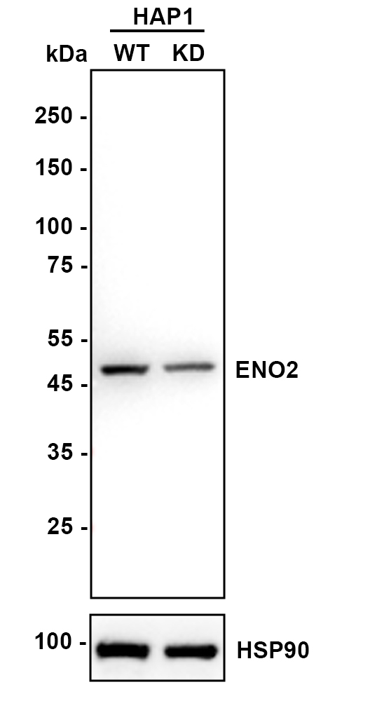
Western blot analysis of NSE on different lysates with Rabbit anti-NSE antibody at 1/5,000 dilution. Lane 1: HAP1-parental cell lysate, Lane 2: HAP1-NSE KD cell lysate, Lysates/proteins at 10 µg/Lane. Exposure time: 6 seconds; 4-20% SDS-PAGE gel. Proteins were transferred to a PVDF membrane and blocked with 5% NFDM/TBST for 1 hour at room temperature. The primary antibody at 1/5,000 dilution was used in K1803 at 4℃ overnight. Goat Anti-Rabbit IgG - HRP Secondary Antibody at 1/50,000 dilution was used for 1 hour at room temperature.IHC
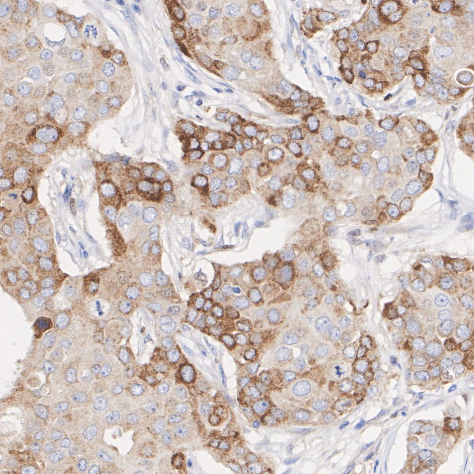
Immunohistochemical analysis of paraffin-embedded human breast cancer tissue with Rabbit anti-NSE antibody at 1/200 dilution. The section was pre-treated using heat mediated antigen retrieval with Tris-EDTA buffer (pH 9.0) for 20 minutes. The tissues were blocked in 1% BSA for 20 minutes at room temperature, washed with ddH2O and PBS, and then probed with the primary antibody at 1/200 dilution for 1 hour at room temperature. The detection was performed using an HRP conjugated compact polymer system. DAB was used as the chromogen. Tissues were counterstained with hematoxylin and mounted with DPX.ICC/IF
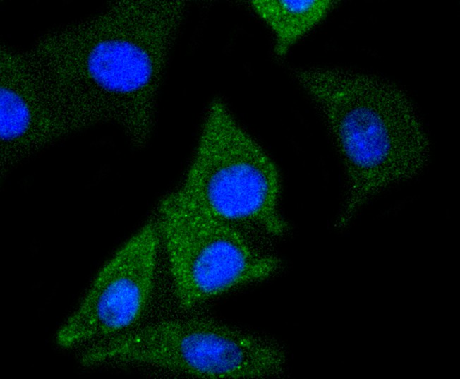
ICC staining of NSE in SH-SY5Y cells (green). Formalin fixed cells were permeabilized with 0.1% Triton X-100 in TBS for 10 minutes at room temperature and blocked with 1% Blocker BSA for 15 minutes at room temperature. Cells were probed with the primary antibody (1/50) for 1 hour at room temperature, washed with PBS. Alexa Fluor®488 Goat anti-Rabbit IgG was used as the secondary antibody at 1/1,000 dilution. The nuclear counter stain is DAPI (blue).| Product Name | NSE Recombinant Rabbit Monoclonal Antibody |
|---|---|
| Antibody Type | Primary Antibodies |
| Product description | d as highly conserved cytoplasmic glycolytic enzymes that may be involved in differentiation. Three isoenzymes have been identified, α Enolase, β Enolase and γ Enolase. α Enolase expression has been detected on most tissues, whereas β Enolase is expressed predominantly in muscle tissue and γ Enolase is detected only in nervous tissue. These isoforms exist as both homodimers and heterodimers, and they play a role in converting phosphoglyceric acid to phosphenolpyruvic acid in the glycolytic pathway. |
| Immunogen | Synthetic peptide within human NSE aa 30-70. |
| Clonality | Monoclonal |
|---|---|
| Isotype | IgG |
| Host Species | Recombinant rabbit |
| Tested Applications | ICC/IFIHCWB |
| WB:1:1000-1:5000 IHC:1:50-1:200 ICC/IF:1:50-1:100 |
|
| Species Reactivity | HumanMouseRatZebrafish |
| Concentration | 1mg/ml |
| Purification | Protein A |
| Alternative Names | 2 phospho D glycerate hydrolyase antibody
2-phospho-D-glycerate hydro-lyase antibody Eno 2 antibody ENO2 antibody ENOG antibody ENOG_HUMAN antibody Enolase 2 (gamma neuronal) antibody Enolase 2 antibody Enolase 2 gamma neuronal antibody Enolase2 antibody Epididymis secretory protein Li 279 antibody Gamma enolase antibody Gamma-enolase antibody HEL S 279 antibody Neural enolase antibody Neuron specific enolase antibody Neuron specific gamma enolase antibody Neuron-specific enolase antibody Neurone specific enolase antibody NSE antibody |
|---|---|
| Molecular Weight(MW) | 47 kDa |
| Cellular Localization | Cytoplasm, Cell membrane. |
WB
Western blot analysis of NSE on different lysates with Rabbit anti-NSE antibody at 1/1,000 dilution. Lane 1: HepG2 cell lysate (10 µg/Lane), Lane 2: HeLa cell lysate (10 µg/Lane), Lane 3: HEK-293 cell lysate (10 µg/Lane), Lane 4: Mouse brain tissue lysate (20 µg/Lane), Lane 5: PC-12 cell lysate (10 µg/Lane), Lane 6: SH-SY5Y cell lysate (10 µg/Lane), Exposure time: 1 minutes; 4-20% SDS-PAGE gel. Proteins were transferred to a PVDF membrane and blocked with 5% NFDM/TBST for 1 hour at room temperature. The primary antibody at 1/1,000 dilution was used in 5% NFDM/TBST at room temperature for 2 hours. Goat Anti-Rabbit IgG - HRP Secondary Antibody at 1:100,000 dilution was used for 1 hour at room temperature.
WB
Western blot analysis of NSE on different lysates with Rabbit anti-NSE antibody at 1/5,000 dilution. Lane 1: HAP1-parental cell lysate, Lane 2: HAP1-NSE KD cell lysate, Lysates/proteins at 10 µg/Lane. Exposure time: 6 seconds; 4-20% SDS-PAGE gel. Proteins were transferred to a PVDF membrane and blocked with 5% NFDM/TBST for 1 hour at room temperature. The primary antibody at 1/5,000 dilution was used in K1803 at 4℃ overnight. Goat Anti-Rabbit IgG - HRP Secondary Antibody at 1/50,000 dilution was used for 1 hour at room temperature.
IHC
Immunohistochemical analysis of paraffin-embedded human breast cancer tissue with Rabbit anti-NSE antibody at 1/200 dilution. The section was pre-treated using heat mediated antigen retrieval with Tris-EDTA buffer (pH 9.0) for 20 minutes. The tissues were blocked in 1% BSA for 20 minutes at room temperature, washed with ddH2O and PBS, and then probed with the primary antibody at 1/200 dilution for 1 hour at room temperature. The detection was performed using an HRP conjugated compact polymer system. DAB was used as the chromogen. Tissues were counterstained with hematoxylin and mounted with DPX.
ICC/IF
ICC staining of NSE in SH-SY5Y cells (green). Formalin fixed cells were permeabilized with 0.1% Triton X-100 in TBS for 10 minutes at room temperature and blocked with 1% Blocker BSA for 15 minutes at room temperature. Cells were probed with the primary antibody (1/50) for 1 hour at room temperature, washed with PBS. Alexa Fluor®488 Goat anti-Rabbit IgG was used as the secondary antibody at 1/1,000 dilution. The nuclear counter stain is DAPI (blue).| Application Notes | WB:1:1000-1:5000 IHC:1:50-1:200 ICC/IF:1:50-1:100 |
|---|
| Form | Liquid |
|---|---|
| Storage Instructions | Store at +4℃ after thawing. Aliquot store at -20℃ or -80℃. Avoid repeated freeze / thaw cycles. |
| Storage Buffer | 1*TBS (pH7.4), 1%BSA, 40%Glycerol. Preservative: 0.05% Sodium Azide. |


