Application
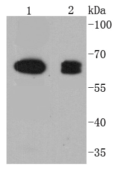
Fig1: Western blot analysis of COX1 on different lysates using anti-COX1 antibody at 1/1,000 dilution. Positive control: Lane 1: C2C12 Lane 2: A431Application
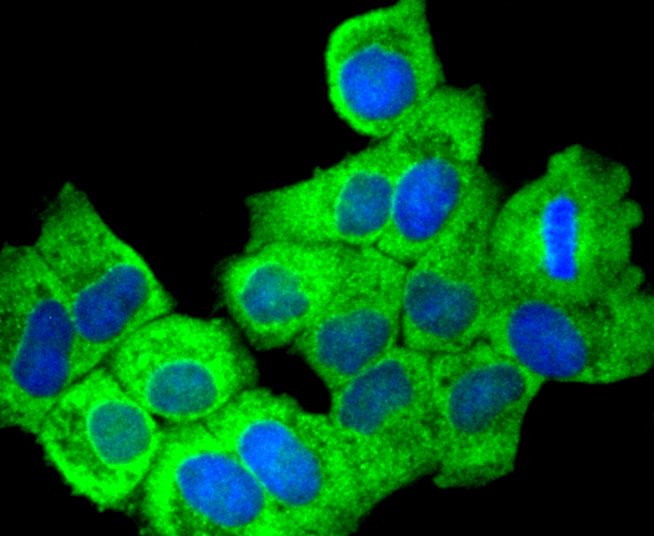
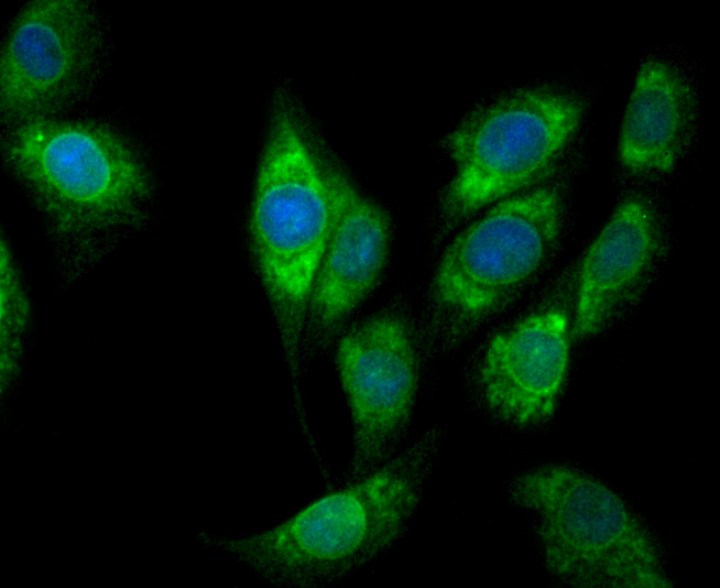
Fig2: ICC staining COX1 in Hela cells (green). The nuclear counter stain is DAPI (blue). Cells were fixed in paraformaldehyde, permeabilised with 0.25% Triton X100/PBS.Application
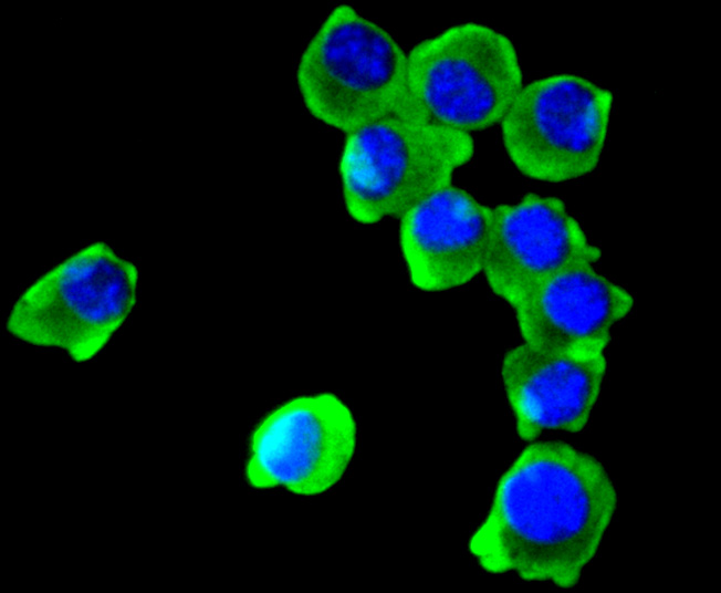
Fig3: ICC staining COX1 in N2A cells (green). The nuclear counter stain is DAPI (blue). Cells were fixed in paraformaldehyde, permeabilised with 0.25% Triton X100/PBS.Application
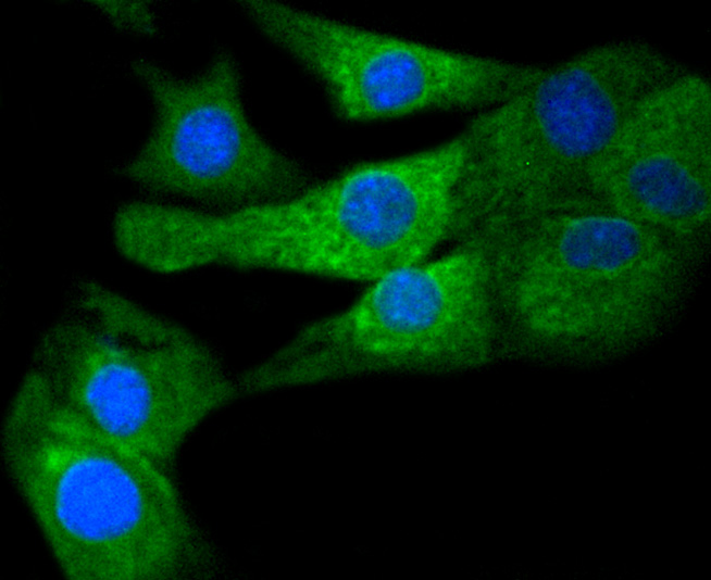
Fig4: ICC staining COX1 in C2C12 cells (green). The nuclear counter stain is DAPI (blue). Cells were fixed in paraformaldehyde, permeabilised with 0.25% Triton X100/PBS.Application
Fig5: ICC staining COX1 in L6 cells (green). The nuclear counter stain is DAPI (blue). Cells were fixed in paraformaldehyde, permeabilised with 0.25% Triton X100/PBS.Application
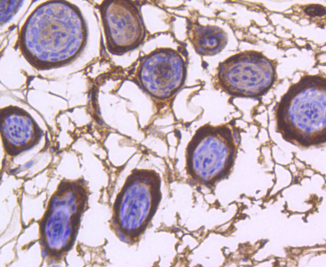
Fig6: Immunohistochemical analysis of paraffin-embedded mouse skin tissue using anti-COX1 antibody. Counter stained with hematoxylin.Application
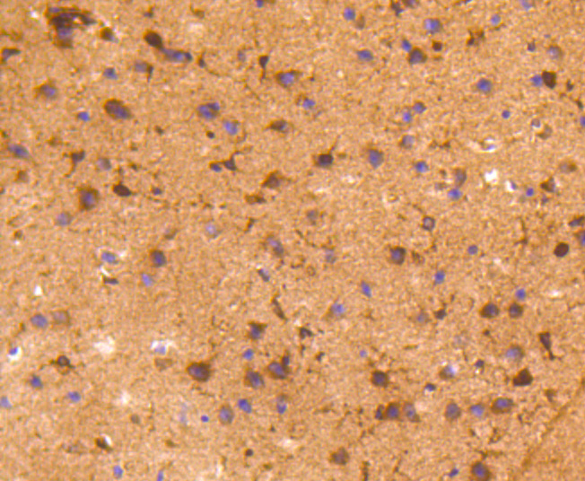
Fig7: Immunohistochemical analysis of paraffin-embedded mouse brain tissue using anti-COX1 antibody. Counter stained with hematoxylin.Application
Fig8: Immunohistochemical analysis of paraffin-embedded human breast cancer tissue using anti-COX1 antibody. Counter stained with hematoxylin.Application
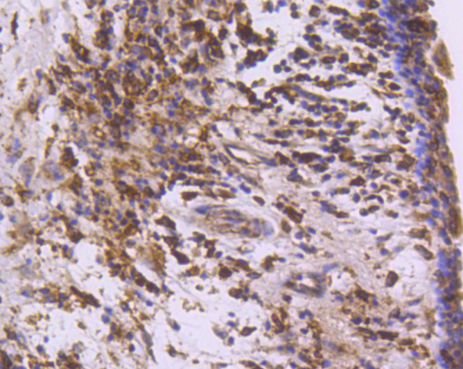
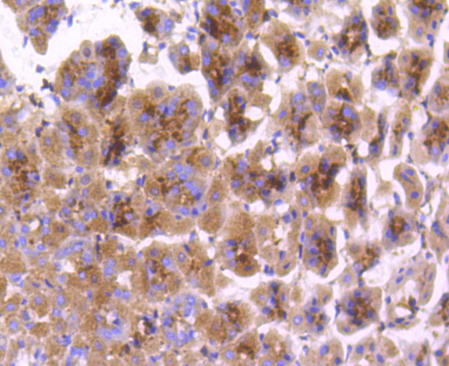
Fig9: Immunohistochemical analysis of paraffin-embedded mouse stomach tissue using anti-COX1 antibody. Counter stained with hematoxylin.Application
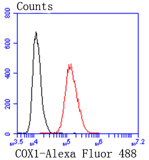
Fig10: Flow cytometric analysis of Hela cells with COX1 antibody at 1/50 dilution (red) compared with an unlabelled control (cells without incubation with primary antibody; black). Alexa Fluor 488-conjugated goat anti rabbit IgG was used as the secondary antibody.| Product Name | COX1/Cyclooxygenase 1 [SC68-05] |
|---|---|
| Antibody Type | Primary Antibodies |
| Antigen Alias | COX 1 antibody COX 3 antibody COX-1 antibody COX1 antibody Cox3 antibody Cyclooxygenase 1 antibody Cyclooxygenase 3, included antibody Cyclooxygenase-1 antibody EC 1.14.99.1 antibody Partial COX1 proteins, included antibody PCOX1 antibody PGG/HS antibody PGH synthase 1 antibody PGH1_HUMAN antibody PGHS-1 antibody PGHS1 antibody PHS 1 antibody PHS1 antibody Prostaglandin G/H synthase 1 antibody Prostaglandin H2 synthase 1 antibody Prostaglandin-endoperoxide synthase 1 (prostaglandin G/H synthase and cyclooxygenase) antibody Prostaglandin-endoperoxide synthase 1 antibody PTGHS antibody PTGS1 antibody |
| Product description | Cytochrome c oxidase subunit I, COX1 (also designated COI, MTCO1 or oxidative phosphorylation (OxPhos) Complex IV, subunit I) is one of three mitochondrial DNA (mtDNA) encoded subunits (MTCO1-3) of respiratory Complex IV. Cytochrome c oxidase is a hetero-oligomeric enzyme composed of 13 subunits localized to the mitochondrial inner membrane and is the terminal enzyme complex of the electron transport chain. Complex IV catalyzes the reduction of molecular oxygen to water. The energy released is used to transport protons across the mitochondrial inner membrane. The resulting electro-chemical gradient is necessary for the synthesis of ATP. Complex IV contains 13 polypeptides; COX1, COX2 and COX3 (MTCO1-3) make up the catalytic core and are encoded by mtDNA while subunits IV, Va, Vb, VIa, VIb, VIc, VIIa, VIIb, VIIc and VIII are nuclear-encoded. |
| Immunogen | recombinant protein |
| Clonality | Monoclonal |
|---|---|
| Isotype | IgG |
| Host Species | Recombinant rabbit |
| Tested Applications | WBICCIHCFCIP |
| WB:1:1,000-1:5,000 ICC:1:100-1:500 IHC:1:50-1:200 FC:1:50-1:100 |
|
| Species Reactivity | HumanMouseRat |
| Concentration | 1mg/ml |
| Alternative Names | COX 1 antibody
COX 3 antibody
COX-1 antibody
COX1 antibody
Cox3 antibody
Cyclooxygenase 1 antibody
Cyclooxygenase 3 included antibody Cyclooxygenase-1 antibody EC 1.14.99.1 antibody Partial COX1 proteins included antibody PCOX1 antibody PGG/HS antibody PGH synthase 1 antibody PGH1_HUMAN antibody PGHS-1 antibody PGHS1 antibody PHS 1 antibody PHS1 antibody Prostaglandin G/H synthase 1 antibody Prostaglandin H2 synthase 1 antibody Prostaglandin-endoperoxide synthase 1 (prostaglandin G/H synthase and cyclooxygenase) antibody Prostaglandin-endoperoxide synthase 1 antibody PTGHS antibody PTGS1 antibody |
|---|---|
| Molecular Weight(MW) | 69 kDa |
| Cellular Localization | Microsome membrane, Endoplasmic reticulum membrane. |
Application
Fig1: Western blot analysis of COX1 on different lysates using anti-COX1 antibody at 1/1,000 dilution. Positive control: Lane 1: C2C12 Lane 2: A431
Application
Fig2: ICC staining COX1 in Hela cells (green). The nuclear counter stain is DAPI (blue). Cells were fixed in paraformaldehyde, permeabilised with 0.25% Triton X100/PBS.
Application
Fig3: ICC staining COX1 in N2A cells (green). The nuclear counter stain is DAPI (blue). Cells were fixed in paraformaldehyde, permeabilised with 0.25% Triton X100/PBS.
Application
Fig4: ICC staining COX1 in C2C12 cells (green). The nuclear counter stain is DAPI (blue). Cells were fixed in paraformaldehyde, permeabilised with 0.25% Triton X100/PBS.
Application
Fig5: ICC staining COX1 in L6 cells (green). The nuclear counter stain is DAPI (blue). Cells were fixed in paraformaldehyde, permeabilised with 0.25% Triton X100/PBS.
Application
Fig6: Immunohistochemical analysis of paraffin-embedded mouse skin tissue using anti-COX1 antibody. Counter stained with hematoxylin.
Application
Fig7: Immunohistochemical analysis of paraffin-embedded mouse brain tissue using anti-COX1 antibody. Counter stained with hematoxylin.
Application
Fig8: Immunohistochemical analysis of paraffin-embedded human breast cancer tissue using anti-COX1 antibody. Counter stained with hematoxylin.
Application
Fig9: Immunohistochemical analysis of paraffin-embedded mouse stomach tissue using anti-COX1 antibody. Counter stained with hematoxylin.
Application
Fig10: Flow cytometric analysis of Hela cells with COX1 antibody at 1/50 dilution (red) compared with an unlabelled control (cells without incubation with primary antibody; black). Alexa Fluor 488-conjugated goat anti rabbit IgG was used as the secondary antibody.| Positive Control | C2C12, A431, Hela, N2A, L6, mouse skin tissue, mouse brain tissue, human breast cancer tissue, mouse stomach tissue. |
|---|---|
| Application Notes | WB:1:1,000-1:5,000 ICC:1:100-1:500 IHC:1:50-1:200 FC:1:50-1:100 |
| Form | Liquid |
|---|---|
| Storage Instructions | Store at +4℃ after thawing. Aliquot store at -20℃ or -80℃. Avoid repeated freeze / thaw cycles. |
| Storage Buffer | 1*TBS (pH7.4), 1%BSA, 40%Glycerol. Preservative: 0.05% Sodium Azide. |


