Application
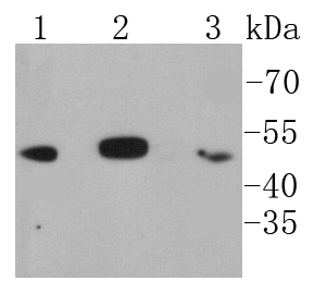
Fig1: Western blot analysis of IKB beta on different lysates using anti-IKB beta antibody at 1/1,000 dilution. Positive control: Lane 1: Jurkat Lane 2: Hela Lane 3: THP-1Application
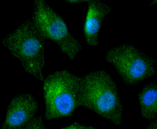
Fig2: ICC staining IKB beta in A549 cells (green). The nuclear counter stain is DAPI (blue). Cells were fixed in paraformaldehyde, permeabilised with 0.25% Triton X100/PBS.Application
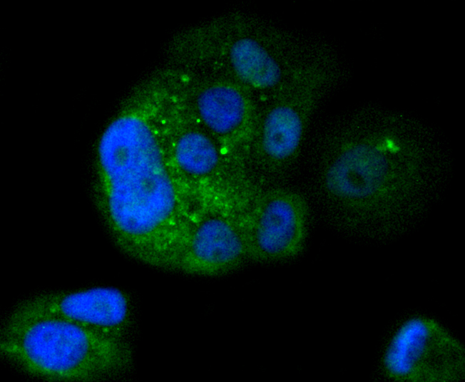
Fig3: ICC staining IKB beta in MCF-7 cells (green). The nuclear counter stain is DAPI (blue). Cells were fixed in paraformaldehyde, permeabilised with 0.25% Triton X100/PBS.Application
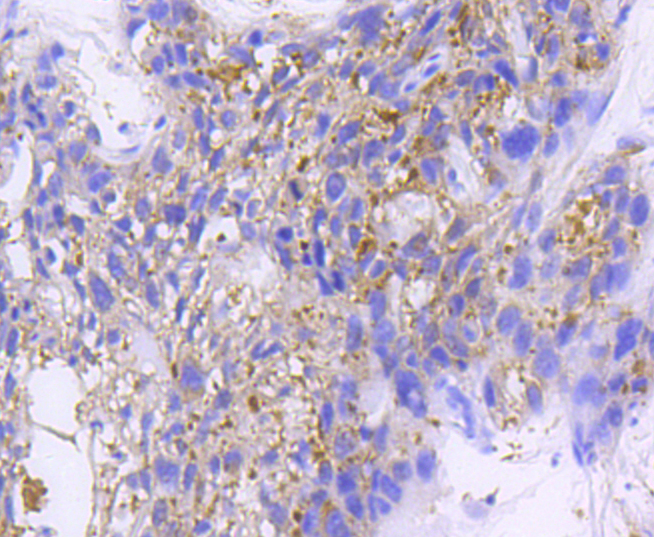
Fig4: Immunohistochemical analysis of paraffin-embedded human breast carcinoma tissue using anti-IKB beta antibody. Counter stained with hematoxylin.| Product Name | IKB beta [SN0648] |
|---|---|
| Antibody Type | Primary Antibodies |
| Antigen Alias | I kappa B beta antibody I-kappa-B-beta antibody IkappaBbeta antibody IKB beta antibody IkB-B antibody IkB-beta antibody IKBB antibody IKBB_HUMAN antibody IkBbeta antibody NF kappa BIB antibody NF-kappa-B inhibitor beta antibody NF-kappa-BIB antibody Nfkbib antibody Thyroid receptor interacting protein 9 antibody Thyroid receptor-interacting protein 9 antibody TR interacting protein 9 antibody TR-interacting protein 9 antibody TRIP-9 antibody TRIP9 antibody |
| Product description | On the basis of both functional and structural considerations, members of the IkB family of proteins can be divided into four groups. The first of these groups, IkB-α, includes the avian protein pp40 and the mammalian MAD-3, both of which inhibit binding of p50-p65 NFkB complex or Rel protein to their cognate binding sites but do not inhibit the binding of p50 homodimer to kB sites, suggesting that the IkB-α family binds to the p65 subunit of p50-p65 heterocomplex through ankyrin repeats. The second member of the IkB family is represented by a protein designated IkB-β. The third group of IkB proteins is represented by IkB-ε, which is identical in sequence with the C-terminal domain of the p110 precursor of NFkB p50 and is expressed predominantly in lymphoid cells. An additional IkB family member, IkB-é, has several phosphorylated forms and is primarily found complexed with Rel A and/or c-Rel. |
| Immunogen | recombinant protein |
| Clonality | Monoclonal |
|---|---|
| Isotype | IgG |
| Host Species | Recombinant rabbit |
| Tested Applications | WBICCIHCIP |
| WB:1:1,000 ICC:1:50-1:200 IHC:1:50-1:200 |
|
| Species Reactivity | Human |
| Concentration | 1mg/ml |
| Alternative Names | I kappa B beta antibody I-kappa-B-beta antibody IkappaBbeta antibody IKB beta antibody IkB-B antibody IkB-beta antibody IKBB antibody IKBB_HUMAN antibody IkBbeta antibody NF kappa BIB antibody NF-kappa-B inhibitor beta antibody NF-kappa-BIB antibody Nfkbib antibody Thyroid receptor interacting protein 9 antibody Thyroid receptor-interacting protein 9 antibody TR interacting protein 9 antibody TR-interacting protein 9 antibody TRIP-9 antibody TRIP9 antibody |
|---|---|
| Molecular Weight(MW) | 48 kDa |
| Cellular Localization | Cytoplasm, Nucleus. |
| SwissProt ID | Q15653 |
|---|
Application
Fig1: Western blot analysis of IKB beta on different lysates using anti-IKB beta antibody at 1/1,000 dilution. Positive control: Lane 1: Jurkat Lane 2: Hela Lane 3: THP-1
Application
Fig2: ICC staining IKB beta in A549 cells (green). The nuclear counter stain is DAPI (blue). Cells were fixed in paraformaldehyde, permeabilised with 0.25% Triton X100/PBS.
Application
Fig3: ICC staining IKB beta in MCF-7 cells (green). The nuclear counter stain is DAPI (blue). Cells were fixed in paraformaldehyde, permeabilised with 0.25% Triton X100/PBS.
Application
Fig4: Immunohistochemical analysis of paraffin-embedded human breast carcinoma tissue using anti-IKB beta antibody. Counter stained with hematoxylin.| Positive Control | A549, MCF-7, Hela, Jurkat, THP-1, human breast carcinoma tissue. |
|---|---|
| Application Notes | WB:1:1,000 ICC:1:50-1:200 IHC:1:50-1:200 |
| Form | Liquid |
|---|---|
| Storage Instructions | Store at +4℃ after thawing. Aliquot store at -20℃ or -80℃. Avoid repeated freeze / thaw cycles. |
| Storage Buffer | 1*TBS (pH7.4), 1%BSA, 40%Glycerol. Preservative: 0.05% Sodium Azide. |


