WB
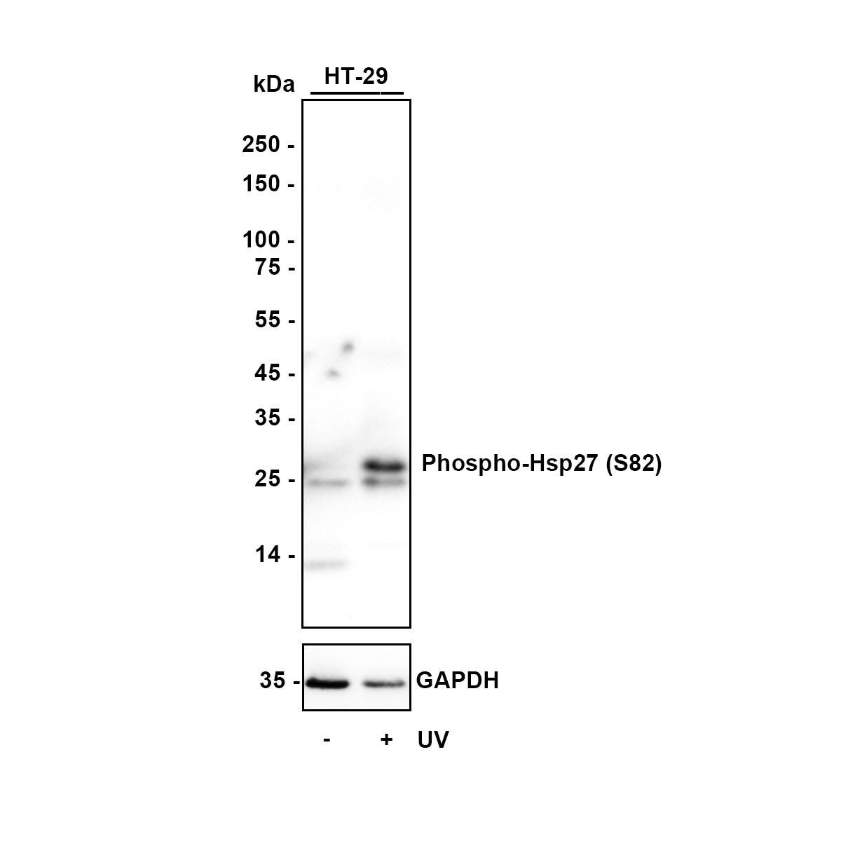
Western blot analysis of Phospho-Hsp27 (S82) on different lysates with Rabbit anti-Phospho-Hsp27 (S82) antibody at 1/1,000 dilution. Lane 1: HT-29 cell lysate, Lane 2: HT-29 treated with UV for 3 hours cell lysate. Lysates/proteins at 20 µg/Lane. Exposure time: 4 seconds; Blocking: 5% NFDM/TBST, 1 hour at room temperature, Primary antibody: 1/1,000 in primary antibody dilution buffer, overnight at 4 ℃ Secondary antibody: Goat anti-Rabbit IgG-HRP, 1/50,000 in 5% NFDM/TBST, 1 hour at room temperature.ICC/IF
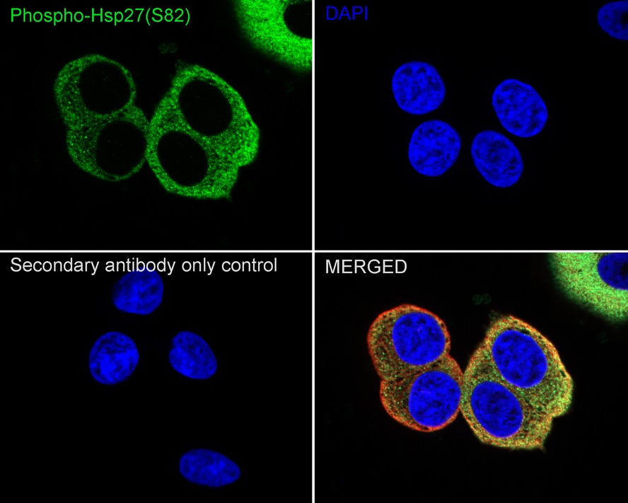
Immunocytochemistry analysis of MCF7 cells labeling Phospho-Hsp27 (S82) with Rabbit anti-Phospho-Hsp27 (S82) antibody at 1/100 dilution. Cells were fixed in 4% paraformaldehyde for 15 minutes at room temperature, permeabilized with 0.1% Triton X-100 in PBS for 15 minutes at room temperature, then blocked with 1% BSA in 10% negative goat serum for 1 hour at room temperature. Cells were then incubated with Rabbit anti-Phospho-Hsp27 (S82) antibody at 1/100 dilution in 1% BSA in PBST overnight at 4 ℃. Goat Anti-Rabbit IgG H&L (488) was used as the secondary antibody at 1/1,000 dilution. PBS instead of the primary antibody was used as the secondary antibody only control. Nuclear DNA was labelled in blue with DAPI. Beta tubulin (red) was stained at 1/100 dilution overnight at +4℃. Goat Anti-Mouse IgG H&L (594) was used as the secondary antibody at 1/1,000 dilution.FC
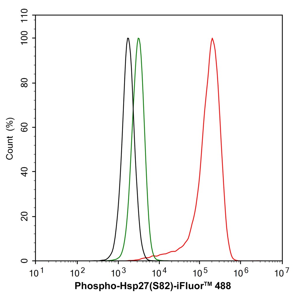
Flow cytometric analysis of MCF7 cells labeling Phospho-Hsp27 (S82). Cells were fixed and permeabilized. Then stained with the primary antibody (1/1,000) (red) compared with Rabbit IgG Isotype Control (green). After incubation of the primary antibody at +4℃ for an hour, the cells were stained with a 488 conjugate-Goat anti-Rabbit IgG Secondary antibody at 1/1,000 dilution for 30 minutes at +4℃. Unlabelled sample was used as a control (cells without incubation with primary antibody; black).| Product Name | Phospho-Hsp27 (S82) Recombinant Rabbit Monoclonal Antibody |
|---|---|
| Antibody Type | Primary Antibodies |
| Product description | HSP 27 is a constitutively expressed cytoplasmic protein that co-localizes to the nucleus upon stress-induced insult. Heat shock, cytokines and hormones are among the factors that stimulate the synthesis of HSP 27. The intracellular concentration of the mammalian heat shock protein, HSP 27, increases several-fold after heat shock and other metabolic stresses, and is closely associated with the acquisition of thermotolerance. MAP kinase-activated protein kinase-2 phosphorylates HSP 27 on Serine residues Ser 15, Ser 78 and Ser 82, which are phosphorylated in vivo in response to growth factors and heat shock. Ser 15, Ser 78 and Ser 82 occur in the sequence motif RXXS, which is recognized by ribosomal protein S6 kinase II. |
| Immunogen | Synthetic phospho-peptide corresponding to residues surrounding Ser82 of Human Hsp27 aa 50-99 / 205. |
| Modification | p-S82 |
| Clonality | Monoclonal |
|---|---|
| Isotype | IgG |
| Host Species | Recombinant rabbit |
| Tested Applications | FCICC/IFWB |
| WB:1:1000 ICC/IF:1:100 FC:1:1000 |
|
| Species Reactivity | HumanMouseRat |
| Concentration | 1mg/ml |
| Purification | Protein A |
| Alternative Names | Heat shock 27kDa protein antibody 28 kDa heat shock protein antibody CMT2F antibody DKFZp586P1322 antibody epididymis secretory protein Li 102 antibody Estrogen regulated 24 kDa protein antibody Estrogen-regulated 24 kDa protein antibody Heat shock 25kDa protein 1 antibody Heat shock 27 kDa protein antibody Heat shock 27kD protein 1 antibody Heat shock 27kDa protein 1 antibody Heat shock 28kDa protein 1 antibody Heat Shock Protein 27 antibody Heat shock protein beta 1 antibody Heat shock protein beta-1 antibody heat shock protein family B (small) member 1 antibody HEL-S-102 antibody HMN2B antibody HS.76067 antibody Hsp 25 antibody HSP 27 antibody Hsp 28 antibody Hsp B1 antibody Hsp25 antibody HSP27 antibody Hsp28 antibody HspB1 antibody HSPB1_HUMAN antibody SRP27 antibody Stress responsive protein 27 antibody Stress-responsive protein 27 antibody |
|---|---|
| Molecular Weight(MW) | 27 kDa |
| Cellular Localization | Cytoplasm, Nucleus. |
WB
Western blot analysis of Phospho-Hsp27 (S82) on different lysates with Rabbit anti-Phospho-Hsp27 (S82) antibody at 1/1,000 dilution. Lane 1: HT-29 cell lysate, Lane 2: HT-29 treated with UV for 3 hours cell lysate. Lysates/proteins at 20 µg/Lane. Exposure time: 4 seconds; Blocking: 5% NFDM/TBST, 1 hour at room temperature, Primary antibody: 1/1,000 in primary antibody dilution buffer, overnight at 4 ℃ Secondary antibody: Goat anti-Rabbit IgG-HRP, 1/50,000 in 5% NFDM/TBST, 1 hour at room temperature.
ICC/IF
Immunocytochemistry analysis of MCF7 cells labeling Phospho-Hsp27 (S82) with Rabbit anti-Phospho-Hsp27 (S82) antibody at 1/100 dilution. Cells were fixed in 4% paraformaldehyde for 15 minutes at room temperature, permeabilized with 0.1% Triton X-100 in PBS for 15 minutes at room temperature, then blocked with 1% BSA in 10% negative goat serum for 1 hour at room temperature. Cells were then incubated with Rabbit anti-Phospho-Hsp27 (S82) antibody at 1/100 dilution in 1% BSA in PBST overnight at 4 ℃. Goat Anti-Rabbit IgG H&L (488) was used as the secondary antibody at 1/1,000 dilution. PBS instead of the primary antibody was used as the secondary antibody only control. Nuclear DNA was labelled in blue with DAPI. Beta tubulin (red) was stained at 1/100 dilution overnight at +4℃. Goat Anti-Mouse IgG H&L (594) was used as the secondary antibody at 1/1,000 dilution.
FC
Flow cytometric analysis of MCF7 cells labeling Phospho-Hsp27 (S82). Cells were fixed and permeabilized. Then stained with the primary antibody (1/1,000) (red) compared with Rabbit IgG Isotype Control (green). After incubation of the primary antibody at +4℃ for an hour, the cells were stained with a 488 conjugate-Goat anti-Rabbit IgG Secondary antibody at 1/1,000 dilution for 30 minutes at +4℃. Unlabelled sample was used as a control (cells without incubation with primary antibody; black).| Application Notes | WB:1:1000 ICC/IF:1:100 FC:1:1000 |
|---|
| Form | Liquid |
|---|---|
| Storage Instructions | Store at +4℃ after thawing. Aliquot store at -20℃ or -80℃. Avoid repeated freeze / thaw cycles. |
| Storage Buffer | 1*TBS (pH7.4), 1%BSA, 40%Glycerol. Preservative: 0.05% Sodium Azide. |


