Application
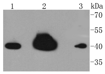
Fig1: Western blot analysis of hnRNP C1+C2 on different lysates using anti-hnRNP C1+C2 antibody at 1/1,000 dilution. Positive control: Lane 1: Hela Lane 2: MCF-7 Lane 3: HepG2Application
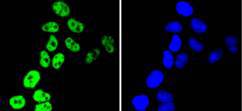
Fig2: ICC staining hnRNP C1+C2 in Hela cells (green). The nuclear counter stain is DAPI (blue). Cells were fixed in paraformaldehyde, permeabilised with 0.25% Triton X100/PBS.Application
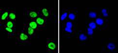
Fig3: ICC staining hnRNP C1+C2 in MCF-7 cells (green). The nuclear counter stain is DAPI (blue). Cells were fixed in paraformaldehyde, permeabilised with 0.25% Triton X100/PBS.Application
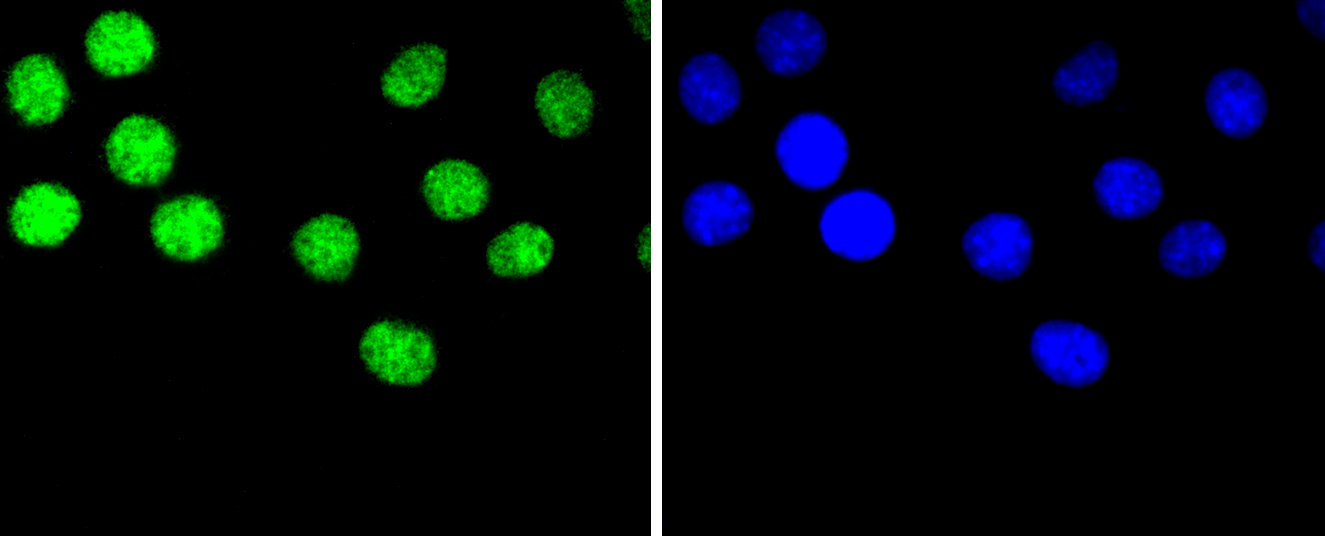
Fig4: ICC staining hnRNP C1+C2 in B16-F1 (green). The nuclear counter stain is DAPI (blue). Cells were fixed in paraformaldehyde, permeabilised with 0.25% Triton X100/PBS.Application
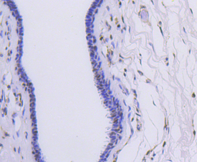
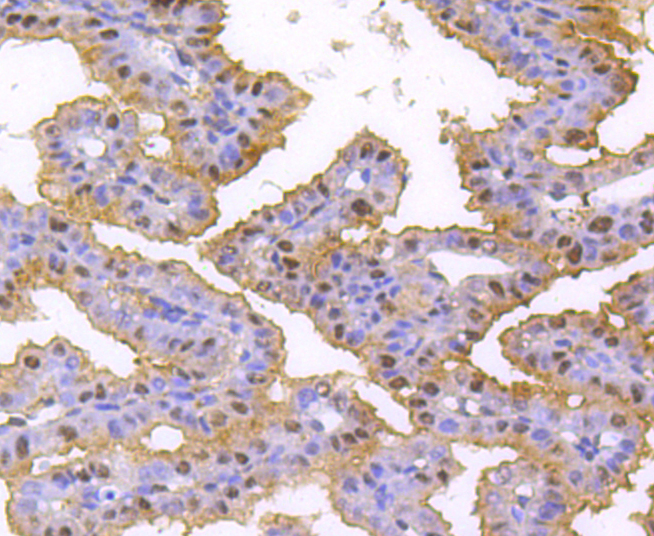
Fig5: Immunohistochemical analysis of paraffin-embedded human breast carcinoma tissue using anti-hnRNP C1+C2 antibody. Counter stained with hematoxylin.Application
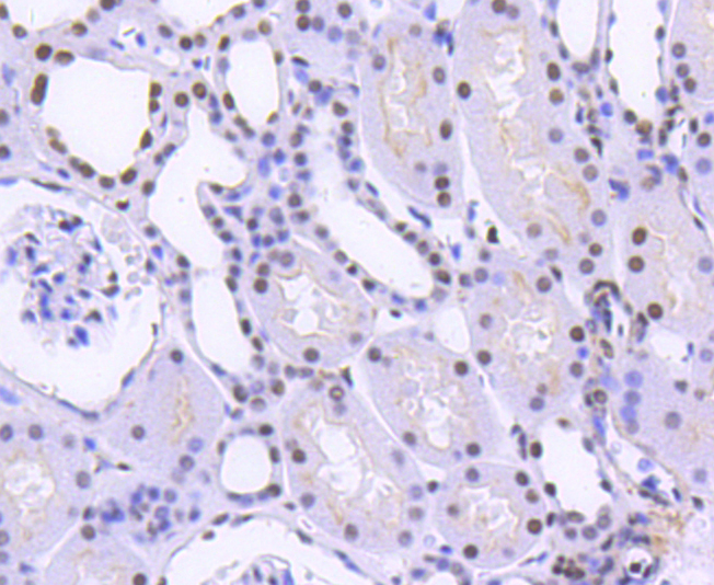
Fig6: Immunohistochemical analysis of paraffin-embedded human kidney tissue using anti-hnRNP C1+C2 antibody. Counter stained with hematoxylin.Application
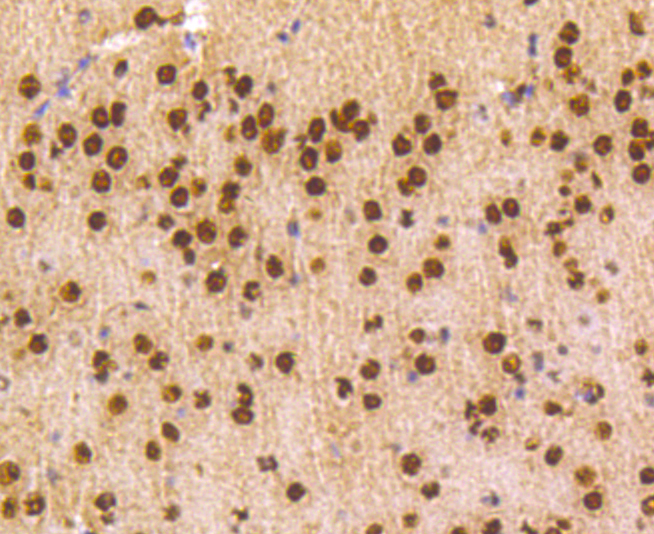
Fig7: Immunohistochemical analysis of paraffin-embedded mouse brain tissue using anti-hnRNP C1+C2 antibody. Counter stained with hematoxylin.Application
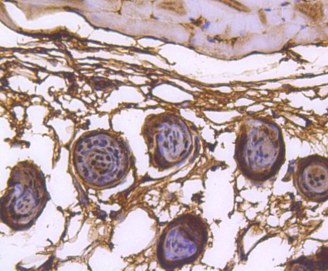
Fig8: Immunohistochemical analysis of paraffin-embedded mouse skin tissue using anti-hnRNP C1+C2 antibody. Counter stained with hematoxylin.Application
Fig9: Immunohistochemical analysis of paraffin-embedded mouse placenta tissue using anti-hnRNP C1+C2 antibody. Counter stained with hematoxylin.Application
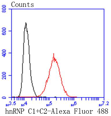
Fig10: Flow cytometric analysis of Hela cells with hnRNP C1+C2 antibody at 1/50 dilution (red) compared with an unlabelled control (cells without incubation with primary antibody; black). Alexa Fluor 488-conjugated goat anti rabbit IgG was used as the secondary antibody| Product Name | hnRNP C1+C2 [SN0652] |
|---|---|
| Antibody Type | Primary Antibodies |
| Antigen Alias | C1 antibody C2 antibody Heterogeneous nuclear ribonucleoprotein C (C1/C2) antibody Heterogeneous nuclear ribonucleoprotein C antibody Heterogeneous nuclear ribonucleoproteins C1/C2 antibody HNRNP antibody hnRNP C1 / hnRNP C2 antibody hnRNP C1/C2 antibody Hnrnpc antibody HNRPC antibody HNRPC_HUMAN antibody MGC104306 antibody MGC105117 antibody MGC117353 antibody MGC131677 antibody Nuclear ribonucleoprotein particle C1 protein antibody Nuclear ribonucleoprotein particle C2 protein antibody SNRPC antibody |
| Product description | Heterogeneous nuclear ribonucleoproteins (hnRNPs) constitute a set of polypeptides that contribute to pre-mRNA processing and transport, and also bind heterogeneous nuclear RNA (hnRNA), which are the transcripts produced by RNA polymerase II. hnRNP complexes are the major constituents of the spliceosome and, in particular, the hnRNP A1 protein is one of the major pre-mRNA/mRNA binding proteins and also one of the most abundant proteins in the nucleus. hnRNP A1 and A2/B1 regulate the processing of pre-mRNA by directly antagonizing the association of various splicing factors and by influencing the splice site selection on pre-mRNA. The majority of hnRNP proteins components are localized to the nucleus; however, some shuttle between the nucleus and the cytoplasm. Most hnRNP proteins, in-cluding hnRNP C1 and C2, contain one or more RNA binding domains and are implicated in the processing of pre-mRNA. hnRNPs F and H are largely related factors that preferentially associate with poly(rG) regions on RNA. Isoforms of these proteins are often generated by alternative processing of the pre-mRNA and by posttranslational modifications such as phosphorylation on serines and threonines and methylation of arginines. |
| Immunogen | recombinant protein |
| Clonality | Monoclonal |
|---|---|
| Isotype | IgG |
| Host Species | Recombinant rabbit |
| Tested Applications | WBICC/IFIHCIPFC |
| WB:1:1,000-1:5,000 ICC:1:100-1:500 IHC:1:50-1:200 FC:1:50-1:100 |
|
| Species Reactivity | HumanMouseRatZebra Fish |
| Concentration | 1mg/ml |
| Alternative Names | C1 antibody C2 antibody Heterogeneous nuclear ribonucleoprotein C (C1/C2) antibody Heterogeneous nuclear ribonucleoprotein C antibody Heterogeneous nuclear ribonucleoproteins C1/C2 antibody HNRNP antibody hnRNP C1 / hnRNP C2 antibody hnRNP C1/C2 antibody Hnrnpc antibody HNRPC antibody HNRPC_HUMAN antibody MGC104306 antibody MGC105117 antibody MGC117353 antibody MGC131677 antibody Nuclear ribonucleoprotein particle C1 protein antibody Nuclear ribonucleoprotein particle C2 protein antibody SNRPC antibody |
|---|---|
| Molecular Weight(MW) | 42 kDa |
| Cellular Localization | Nucleus. |
Application
Fig1: Western blot analysis of hnRNP C1+C2 on different lysates using anti-hnRNP C1+C2 antibody at 1/1,000 dilution. Positive control: Lane 1: Hela Lane 2: MCF-7 Lane 3: HepG2
Application
Fig2: ICC staining hnRNP C1+C2 in Hela cells (green). The nuclear counter stain is DAPI (blue). Cells were fixed in paraformaldehyde, permeabilised with 0.25% Triton X100/PBS.
Application
Fig3: ICC staining hnRNP C1+C2 in MCF-7 cells (green). The nuclear counter stain is DAPI (blue). Cells were fixed in paraformaldehyde, permeabilised with 0.25% Triton X100/PBS.
Application
Fig4: ICC staining hnRNP C1+C2 in B16-F1 (green). The nuclear counter stain is DAPI (blue). Cells were fixed in paraformaldehyde, permeabilised with 0.25% Triton X100/PBS.
Application
Fig5: Immunohistochemical analysis of paraffin-embedded human breast carcinoma tissue using anti-hnRNP C1+C2 antibody. Counter stained with hematoxylin.
Application
Fig6: Immunohistochemical analysis of paraffin-embedded human kidney tissue using anti-hnRNP C1+C2 antibody. Counter stained with hematoxylin.
Application
Fig7: Immunohistochemical analysis of paraffin-embedded mouse brain tissue using anti-hnRNP C1+C2 antibody. Counter stained with hematoxylin.
Application
Fig8: Immunohistochemical analysis of paraffin-embedded mouse skin tissue using anti-hnRNP C1+C2 antibody. Counter stained with hematoxylin.
Application
Fig9: Immunohistochemical analysis of paraffin-embedded mouse placenta tissue using anti-hnRNP C1+C2 antibody. Counter stained with hematoxylin.
Application
Fig10: Flow cytometric analysis of Hela cells with hnRNP C1+C2 antibody at 1/50 dilution (red) compared with an unlabelled control (cells without incubation with primary antibody; black). Alexa Fluor 488-conjugated goat anti rabbit IgG was used as the secondary antibody| Positive Control | B16-F1, MCF-7, Hela, HepG2, mouse skin tissue, human breast carcinoma tissue, mouse placenta tissue, human kidney tissue, mouse brain tissue. |
|---|---|
| Application Notes | WB:1:1,000-1:5,000 ICC:1:100-1:500 IHC:1:50-1:200 FC:1:50-1:100 |
| Form | Liquid |
|---|---|
| Storage Instructions | Store at +4℃ after thawing. Aliquot store at -20℃ or -80℃. Avoid repeated freeze / thaw cycles. |
| Storage Buffer | 1*TBS (pH7.4), 1%BSA, 40%Glycerol. Preservative: 0.05% Sodium Azide. |


