Application
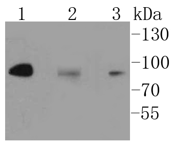
Fig1: Western blot analysis of IKK alpha+IKK beta on different lysates using anti-IKK alpha+IKK beta antibody at 1/1,000 dilution. Positive control: Lane 1: Hela Lane 2: Daudi Lane 3: A431Application
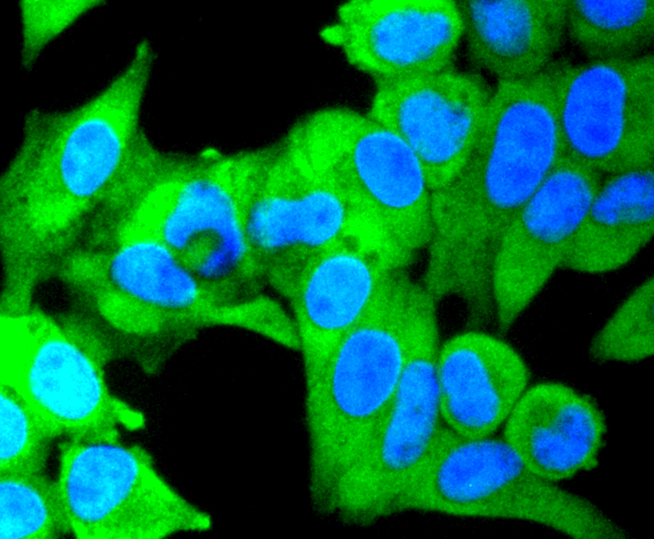
Fig2: ICC staining IKK alpha+IKK beta in Hela cells (green). The nuclear counter stain is DAPI (blue). Cells were fixed in paraformaldehyde, permeabilised with 0.25% Triton X100/PBS.Application
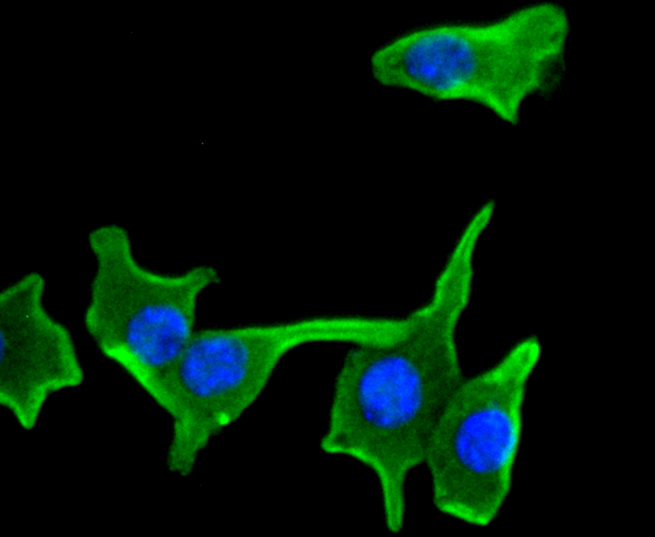
Fig3: ICC staining IKK alpha+IKK beta in B-6F1 cells (green). The nuclear counter stain is DAPI (blue). Cells were fixed in paraformaldehyde, permeabilised with 0.25% Triton X100/PBS.Application
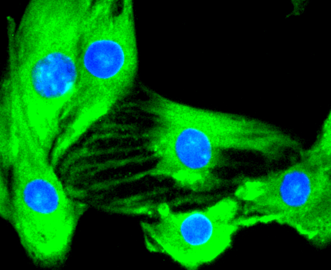
Fig4: ICC staining IKK alpha+IKK beta in C2C12 cells (green). The nuclear counter stain is DAPI (blue). Cells were fixed in paraformaldehyde, permeabilised with 0.25% Triton X100/PBS.| Product Name | IKK alpha + IKK beta [SN63-02] |
|---|---|
| Antibody Type | Primary Antibodies |
| Antigen Alias | CHUK antibody Conserved helix loop helix ubiquitous kinase antibody I kappa B kinase 1 antibody I kappa B kinase 2 antibody I Kappa B kinase alpha antibody I Kappa B kinase beta antibody IkB kinase alpha subunit antibody IkBKA antibody IkBKB antibody IKK a kinase antibody IKK alpha antibody IKK beta antibody IKK1 antibody IKK2 antibody IKKA antibody IKKB antibody IMD15 antibody Inhibitor of kappa light polypeptide gene enhancer in B cells kinase beta antibody Inhibitor of nuclear factor kappa-B kinase subunit alpha antibody Inhibitor of nuclear factor kappa-B kinase subunit beta antibody NFKBIKA antibody NFKBIKB antibody Nuclear factor NF kappa B inhibitor kinase beta antibody Nuclear factor NFkappaB inhibitor kinase alpha antibody TCF 16 antibody TCF16 antibody Transcription factor 16 antibody |
| Product description | The transcription factor NFkB is retained in the cytoplasm in an inactive form by the inhibitory protein IkB. Activation of NFkB requires that IkB be phosphorylated on specific serine residues, which results in targeted degradation of IkB. IkB kinase a (IKKa), previously designated CHUK, interacts with IkB-a and specifically phosphorylates IkB-a on Ser 32 and 36, the sites that trigger its degradation. IKKa appears to be critical for NFkB activation in response to proinflammatory cytokines. Phosphorylation of IkB by IKKa is stimulated by the NFkB inducing kinase (NIK), which itself is a central regulator for NFkB activation in response to TNF and IL-1. The functional IKK complex contains three subunits, IKKa, IKKb and IKKg (also designated NEMO), and each appear to make essential contributions to IkB phosphorylation. |
| Immunogen | recombinant protein |
| Clonality | Monoclonal |
|---|---|
| Isotype | IgG |
| Host Species | Recombinant rabbit |
| Tested Applications | WBICC/IFIP |
| WB:1:1,000 ICC:1:100-1:500 |
|
| Species Reactivity | HumanMouseRat |
| Concentration | 1mg/ml |
| Alternative Names | CHUK antibody Conserved helix loop helix ubiquitous kinase antibody I kappa B kinase 1 antibody I kappa B kinase 2 antibody I Kappa B kinase alpha antibody I Kappa B kinase beta antibody IkB kinase alpha subunit antibody IkBKA antibody IkBKB antibody IKK a kinase antibody IKK alpha antibody IKK beta antibody IKK1 antibody IKK2 antibody IKKA antibody IKKB antibody IMD15 antibody Inhibitor of kappa light polypeptide gene enhancer in B cells kinase beta antibody Inhibitor of nuclear factor kappa-B kinase subunit alpha antibody Inhibitor of nuclear factor kappa-B kinase subunit beta antibody NFKBIKA antibody NFKBIKB antibody Nuclear factor NF kappa B inhibitor kinase beta antibody Nuclear factor NFkappaB inhibitor kinase alpha antibody TCF 16 antibody TCF16 antibody Transcription factor 16 antibody |
|---|---|
| Molecular Weight(MW) | 85/87 kDa |
| Cellular Localization | Cytoplasm, Nucleus, Membrane raft. |
Application
Fig1: Western blot analysis of IKK alpha+IKK beta on different lysates using anti-IKK alpha+IKK beta antibody at 1/1,000 dilution. Positive control: Lane 1: Hela Lane 2: Daudi Lane 3: A431
Application
Fig2: ICC staining IKK alpha+IKK beta in Hela cells (green). The nuclear counter stain is DAPI (blue). Cells were fixed in paraformaldehyde, permeabilised with 0.25% Triton X100/PBS.
Application
Fig3: ICC staining IKK alpha+IKK beta in B-6F1 cells (green). The nuclear counter stain is DAPI (blue). Cells were fixed in paraformaldehyde, permeabilised with 0.25% Triton X100/PBS.
Application
Fig4: ICC staining IKK alpha+IKK beta in C2C12 cells (green). The nuclear counter stain is DAPI (blue). Cells were fixed in paraformaldehyde, permeabilised with 0.25% Triton X100/PBS.| Positive Control | A431, Daudi, Hela, B-6F1, C2C12. |
|---|---|
| Application Notes | WB:1:1,000 ICC:1:100-1:500 |
| Form | Liquid |
|---|---|
| Storage Instructions | Store at +4℃ after thawing. Aliquot store at -20℃ or -80℃. Avoid repeated freeze / thaw cycles. |
| Storage Buffer | 1*TBS (pH7.4), 1%BSA, 40%Glycerol. Preservative: 0.05% Sodium Azide. |


