WB
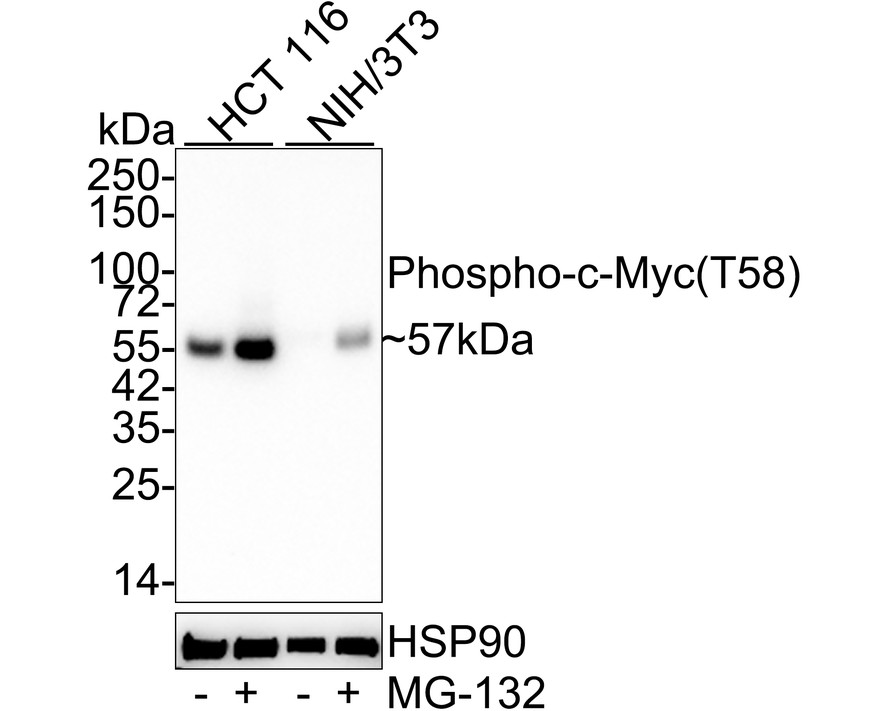
Western blot analysis of Phospho-c-Myc (T58) on different lysates with Rabbit anti-Phospho-c-Myc (T58) antibody at 1/1,000 dilution. Lane 1: HCT 116 cell lysate, Lane 2: HCT 116 treated with 25μM MG-132 for 4 hours cell lysate, Lane 3: NIH/3T3 cell lysate, Lane 4: NIH/3T3 treated with 10μM MG-132 for 8 hours cell lysate, Lysates/proteins at 20 µg/Lane. Exposure time: 2 minutes 18 seconds; 4-20% SDS-PAGE gel. Proteins were transferred to a PVDF membrane and blocked with 5% NFDM/TBST for 1 hour at room temperature. The primary antibody at 1/1,000 dilution was used in 5% NFDM/TBST at 4℃ overnight. Goat Anti-Rabbit IgG - HRP Secondary Antibody at 1/50,000 dilution was used for 1 hour at room temperature.ICC/IF
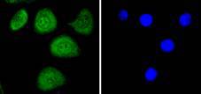
ICC staining phospho-c-Myc(T58) in SKOV-3 cells (green). The nuclear counter stain is DAPI (blue). Cells were fixed in paraformaldehyde, permeabilised with 0.25% Triton X100/PBS.FC
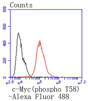
Flow cytometric analysis of MCF-7 cells with phospho-c-Myc(T58) antibody at 1/50 dilution (red) compared with an unlabelled control (cells without incubation with primary antibody; black). Alexa Fluor 488-conjugated goat anti rabbit IgG was used as the secondary antibodyIHC
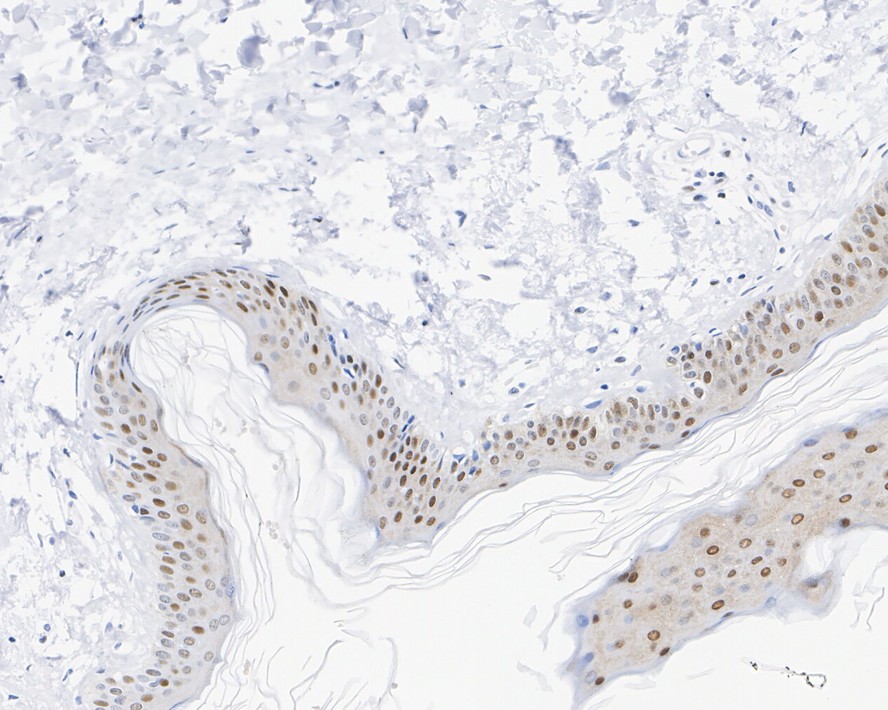
Immunohistochemical analysis of paraffin-embedded human skin tissue with Rabbit anti-Phospho-c-Myc (T58) antibody at 1/500 dilution. The section was pre-treated using heat mediated antigen retrieval with sodium citrate buffer (pH 6.0) for 2 minutes. The tissues were blocked in 1% BSA for 20 minutes at room temperature, washed with ddH2O and PBS, and then probed with the primary antibody at 1/500 dilution for 1 hour at room temperature. The detection was performed using an HRP conjugated compact polymer system. DAB was used as the chromogen. Tissues were counterstained with hematoxylin and mounted with DPX.| Product Name | Phospho-c-Myc (T58) Recombinant Rabbit Monoclonal Antibody |
|---|---|
| Antibody Type | Primary Antibodies |
| Product description | c-Myc-, N-Myc- and L-Myc-encoded proteins function in cell proliferation, differentiation and neoplastic disease. Myc proteins are nuclear proteins with relatively short half lives. Amplification of the c-Myc gene has been found in several types of human tumors including lung, breast and colon carcinomas, while the N-Myc gene has been found amplified in neuroblastomas. The L-Myc gene has been reported to be amplified and expressed at high level in human small cell lung carcinomas. The presence of three sequence motifs in the c-Myc COOH terminus, including the leucine zipper, the helix-loop-helix and a basic region provided initial evidence for a sequence-specific binding function. A basic region helix-loop-helix leucine zipper motif (bHLH-Zip) protein, designated Max, specifically associates with c-Myc, N-Myc and L-Myc proteins. The Myc-Max complex binds to DNA in a sequence-specific manner under conditions where neither Max nor Myc exhibit appreciable binding. Max can also form heterodimers with at least two additional bHLH-Zip proteins, Mad and Mxi1, and Mad-Max dimers have been shown to repress transcription through interaction with mSin3. |
| Immunogen | Synthetic phospho-peptide corresponding to residues surrounding Thr58 of Human Myc (P01106-1). |
| Modification | p-T58 |
| Clonality | monoclonal |
|---|---|
| Isotype | IgG |
| Host Species | Recombinant rabbit |
| Tested Applications | FCICC/IFIHCWB |
| WB:1:1000 FC:1:50-1:100 ICC/IF:1:50-1:200 IHC:1:500 |
|
| Species Reactivity | HumanMouseRat |
| Concentration | 1mg/ml |
| Purification | Protein A |
| Alternative Names | Avian myelocytomatosis viral oncogene homolog antibody bHLHe39 antibody c Myc antibody Class E basic helix-loop-helix protein 39 antibody MRTL antibody Myc antibody Myc protein antibody Myc proto oncogene protein antibody Myc proto-oncogene protein antibody myc related translation/localization regulatory factor antibody MYC_HUMAN antibody Myc2 antibody MYCC antibody Niard antibody Nird antibody Proto-oncogene c-Myc antibody Transcription factor p64 antibody v myc avian myelocytomatosis viral oncogene homolog antibody v myc myelocytomatosis viral oncogene homolog antibody |
|---|---|
| Molecular Weight(MW) | 49kDa(Observed band size: 57kDa) |
| Cellular Localization | Nucleus. |
| SwissProt ID | P01106 |
|---|
WB
Western blot analysis of Phospho-c-Myc (T58) on different lysates with Rabbit anti-Phospho-c-Myc (T58) antibody at 1/1,000 dilution. Lane 1: HCT 116 cell lysate, Lane 2: HCT 116 treated with 25μM MG-132 for 4 hours cell lysate, Lane 3: NIH/3T3 cell lysate, Lane 4: NIH/3T3 treated with 10μM MG-132 for 8 hours cell lysate, Lysates/proteins at 20 µg/Lane. Exposure time: 2 minutes 18 seconds; 4-20% SDS-PAGE gel. Proteins were transferred to a PVDF membrane and blocked with 5% NFDM/TBST for 1 hour at room temperature. The primary antibody at 1/1,000 dilution was used in 5% NFDM/TBST at 4℃ overnight. Goat Anti-Rabbit IgG - HRP Secondary Antibody at 1/50,000 dilution was used for 1 hour at room temperature.
ICC/IF
ICC staining phospho-c-Myc(T58) in SKOV-3 cells (green). The nuclear counter stain is DAPI (blue). Cells were fixed in paraformaldehyde, permeabilised with 0.25% Triton X100/PBS.
FC
Flow cytometric analysis of MCF-7 cells with phospho-c-Myc(T58) antibody at 1/50 dilution (red) compared with an unlabelled control (cells without incubation with primary antibody; black). Alexa Fluor 488-conjugated goat anti rabbit IgG was used as the secondary antibody
IHC
Immunohistochemical analysis of paraffin-embedded human skin tissue with Rabbit anti-Phospho-c-Myc (T58) antibody at 1/500 dilution. The section was pre-treated using heat mediated antigen retrieval with sodium citrate buffer (pH 6.0) for 2 minutes. The tissues were blocked in 1% BSA for 20 minutes at room temperature, washed with ddH2O and PBS, and then probed with the primary antibody at 1/500 dilution for 1 hour at room temperature. The detection was performed using an HRP conjugated compact polymer system. DAB was used as the chromogen. Tissues were counterstained with hematoxylin and mounted with DPX.| Application Notes | WB:1:1000 FC:1:50-1:100 ICC/IF:1:50-1:200 IHC:1:500 |
|---|
| Form | Liquid |
|---|---|
| Storage Instructions | Store at +4℃ after thawing. Aliquot store at -20℃ or -80℃. Avoid repeated freeze / thaw cycles. |
| Storage Buffer | 1*TBS (pH7.4), 1%BSA, 40%Glycerol. Preservative: 0.05% Sodium Azide. |


