WB
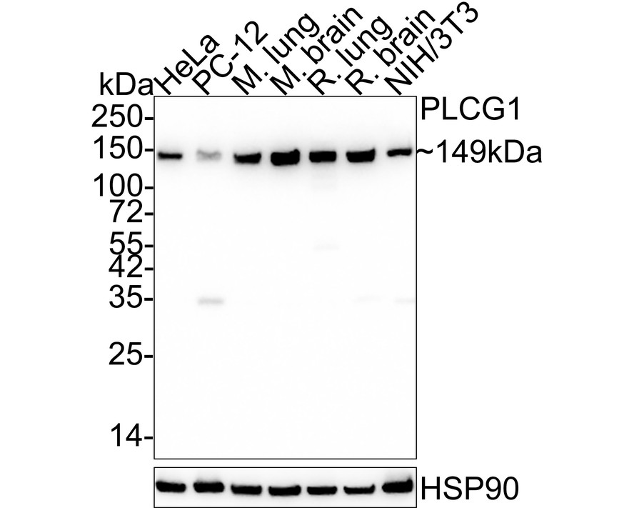
Western blot analysis of PLC-gamma-1 on different lysates with Rabbit anti-PLC-gamma-1 antibody at 1/1,000 dilution. Lane 1: HeLa cell lysate (20 µg/Lane), Lane 2: PC-12 cell lysate (20 µg/Lane), Lane 3: Mouse lung tissue lysate (40 µg/Lane), Lane 4: Mouse brain tissue lysate (40 µg/Lane), Lane 5: Rat lung tissue lysate (40 µg/Lane), Lane 6: Rat brain tissue lysate (40 µg/Lane), Lane 7: NIH/3T3 cell lysate (20 µg/Lane), Exposure time: 50 seconds; 4-20% SDS-PAGE gel. Proteins were transferred to a PVDF membrane and blocked with 5% NFDM/TBST for 1 hour at room temperature. The primary antibody at 1/1,000 dilution was used in 5% NFDM/TBST at 4℃ overnight. Goat Anti-Rabbit IgG - HRP Secondary Antibody at 1/50,000 dilution was used for 1 hour at room temperature.IHC
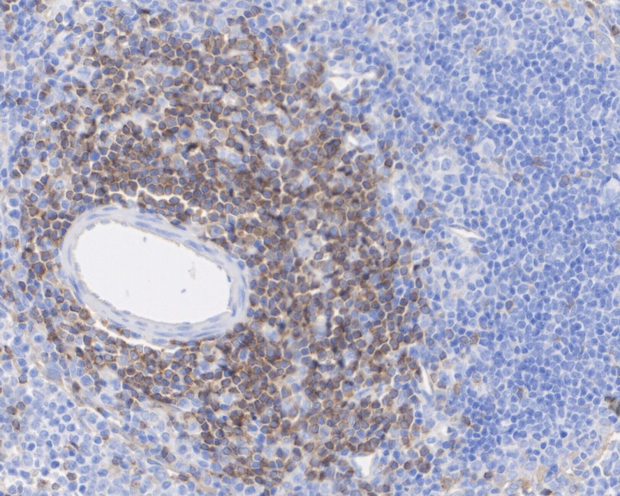
Immunohistochemical analysis of paraffin-embedded rat spleen tissue with Rabbit anti-PLC-gamma-1 antibody at 1/200 dilution. The section was pre-treated using heat mediated antigen retrieval with Tris-EDTA buffer (pH 9.0) for 20 minutes. The tissues were blocked in 1% BSA for 20 minutes at room temperature, washed with ddH2O and PBS, and then probed with the primary antibody at 1/200 dilution for 1 hour at room temperature. The detection was performed using an HRP conjugated compact polymer system. DAB was used as the chromogen. Tissues were counterstained with hematoxylin and mounted with DPX.IHC
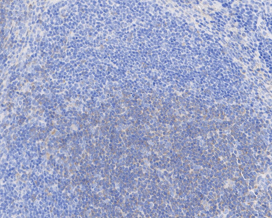
Immunohistochemical analysis of paraffin-embedded mouse spleen tissue with Rabbit anti-PLC-gamma-1 antibody at 1/200 dilution. The section was pre-treated using heat mediated antigen retrieval with Tris-EDTA buffer (pH 9.0) for 20 minutes. The tissues were blocked in 1% BSA for 20 minutes at room temperature, washed with ddH2O and PBS, and then probed with the primary antibody at 1/200 dilution for 1 hour at room temperature. The detection was performed using an HRP conjugated compact polymer system. DAB was used as the chromogen. Tissues were counterstained with hematoxylin and mounted with DPX.ICC/IF
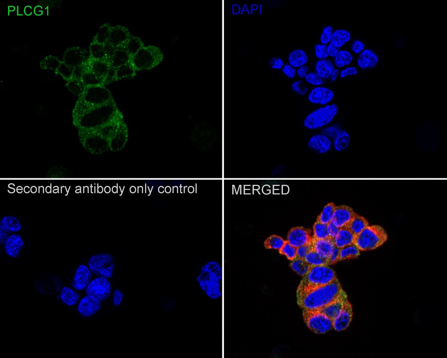
Immunocytochemistry analysis of PC-12 cells labeling PLC-gamma-1 with Rabbit anti-PLC-gamma-1 antibody at 1/50 dilution. Cells were fixed in 4% paraformaldehyde for 20 minutes at room temperature, permeabilized with 0.1% Triton X-100 in PBS for 5 minutes at room temperature, then blocked with 1% BSA in 10% negative goat serum for 1 hour at room temperature. Cells were then incubated with Rabbit anti-PLC-gamma-1 antibody at 1/50 dilution in 1% BSA in PBST overnight at 4 ℃. Goat Anti-Rabbit IgG H&L (488) was used as the secondary antibody at 1/1,000 dilution. PBS instead of the primary antibody was used as the secondary antibody only control. Nuclear DNA was labelled in blue with DAPI. Beta tubulin (red) was stained at 1/100 dilution overnight at +4℃. Goat Anti-Mouse IgG H&L (594) was used as the secondary antibody at 1/1,000 dilution.| Product Name | PLC-gamma-1 Recombinant Rabbit Monoclonal Antibody |
|---|---|
| Antibody Type | Primary Antibodies |
| Product description | Phosphoinositide-specific phospholipase C (PLC) plays a crucial role in the initiation of receptor mediated signal transduction through the generation of the two second messengers, inositol 1,4,5-triphosphate and diacylglycerol from phosphatidylinositol 4,5-bisphosphate. There are many mammalian PLC isozymes, including PLC β1, PLC β2, PLC β3, PLC β4, PLC γ1, PLC γ2, PLC δ1, PLC δ2 and PLCε. PLC γ1 is widely distributed in bronchiolar epithelium, type I and II pneumocytes and fibroblasts of the interstitial tissue. Actin-regulatory protein Villin is tyrosine phosphorylated and associates with PLC γ1 in the brush border of intestinal epithelial cells. Villin regulates PLC γ1 activity by modifying its own ability to bind phosphatidylinositol 4,5-biphosphate. PLC γ1 binds Integrin α1/β1 and modulates Integrin α1/β-specific adhesion. PLC γ1 and Ca2+ play a direct role in VEGF-regulated endothelial growth, however this signaling pathway is not linked to FGF-mediated effects in primary endothelial cells. PLC γ1 is rapidly activated in response to growth factor stimulation and plays an important role in regulating cell proliferation and differentiation. It may also have a protective function during cellular response to oxidative stress. |
| Immunogen | Recombinant protein within Human PLC-gamma-1 aa 690-852 / 1,290. |
| Clonality | Monoclonal |
|---|---|
| Isotype | IgG |
| Host Species | Recombinant rabbit |
| Tested Applications | ICC/IFIHCWB |
| WB:1:1000 IHC:1:200 ICC/IF:1:50-1:200 |
|
| Species Reactivity | HumanMouseRat |
| Concentration | 1mg/ml |
| Purification | Protein A |
| Alternative Names | 1 phosphatidyl D myo inositol 4 5 bisphosphate antibody
1 phosphatidylinositol 4 5 bisphosphate phosphodiesterase gamma 1 antibody
1-phosphatidylinositol-4 5-bisphosphate phosphodiesterase gamma-1 antibody Inositoltrisphosphohydrolase antibody Monophosphatidylinositol phosphodiesterase antibody NCKAP3 antibody Phosphatidylinositol phospholipase C antibody Phosphoinositidase C antibody Phosphoinositide phospholipase C antibody Phosphoinositide phospholipase C-gamma-1 antibody Phospholipase C 148 antibody Phospholipase C gamma 1 antibody Phospholipase C-gamma-1 antibody Phospholipase C-II antibody PLC gamma 1 antibody PLC II antibody PLC-148 antibody PLC-gamma-1 antibody PLC-II antibody PLC1 antibody PLC148 antibody Plcg1 antibody PLCG1_HUMAN antibody PLCgamma1 antibody |
|---|---|
| Molecular Weight(MW) | 149 kDa |
| Cellular Localization | Cell projection. |
WB
Western blot analysis of PLC-gamma-1 on different lysates with Rabbit anti-PLC-gamma-1 antibody at 1/1,000 dilution. Lane 1: HeLa cell lysate (20 µg/Lane), Lane 2: PC-12 cell lysate (20 µg/Lane), Lane 3: Mouse lung tissue lysate (40 µg/Lane), Lane 4: Mouse brain tissue lysate (40 µg/Lane), Lane 5: Rat lung tissue lysate (40 µg/Lane), Lane 6: Rat brain tissue lysate (40 µg/Lane), Lane 7: NIH/3T3 cell lysate (20 µg/Lane), Exposure time: 50 seconds; 4-20% SDS-PAGE gel. Proteins were transferred to a PVDF membrane and blocked with 5% NFDM/TBST for 1 hour at room temperature. The primary antibody at 1/1,000 dilution was used in 5% NFDM/TBST at 4℃ overnight. Goat Anti-Rabbit IgG - HRP Secondary Antibody at 1/50,000 dilution was used for 1 hour at room temperature.
IHC
Immunohistochemical analysis of paraffin-embedded rat spleen tissue with Rabbit anti-PLC-gamma-1 antibody at 1/200 dilution. The section was pre-treated using heat mediated antigen retrieval with Tris-EDTA buffer (pH 9.0) for 20 minutes. The tissues were blocked in 1% BSA for 20 minutes at room temperature, washed with ddH2O and PBS, and then probed with the primary antibody at 1/200 dilution for 1 hour at room temperature. The detection was performed using an HRP conjugated compact polymer system. DAB was used as the chromogen. Tissues were counterstained with hematoxylin and mounted with DPX.
IHC
Immunohistochemical analysis of paraffin-embedded mouse spleen tissue with Rabbit anti-PLC-gamma-1 antibody at 1/200 dilution. The section was pre-treated using heat mediated antigen retrieval with Tris-EDTA buffer (pH 9.0) for 20 minutes. The tissues were blocked in 1% BSA for 20 minutes at room temperature, washed with ddH2O and PBS, and then probed with the primary antibody at 1/200 dilution for 1 hour at room temperature. The detection was performed using an HRP conjugated compact polymer system. DAB was used as the chromogen. Tissues were counterstained with hematoxylin and mounted with DPX.
ICC/IF
Immunocytochemistry analysis of PC-12 cells labeling PLC-gamma-1 with Rabbit anti-PLC-gamma-1 antibody at 1/50 dilution. Cells were fixed in 4% paraformaldehyde for 20 minutes at room temperature, permeabilized with 0.1% Triton X-100 in PBS for 5 minutes at room temperature, then blocked with 1% BSA in 10% negative goat serum for 1 hour at room temperature. Cells were then incubated with Rabbit anti-PLC-gamma-1 antibody at 1/50 dilution in 1% BSA in PBST overnight at 4 ℃. Goat Anti-Rabbit IgG H&L (488) was used as the secondary antibody at 1/1,000 dilution. PBS instead of the primary antibody was used as the secondary antibody only control. Nuclear DNA was labelled in blue with DAPI. Beta tubulin (red) was stained at 1/100 dilution overnight at +4℃. Goat Anti-Mouse IgG H&L (594) was used as the secondary antibody at 1/1,000 dilution.| Application Notes | WB:1:1000 IHC:1:200 ICC/IF:1:50-1:200 |
|---|
| Form | Liquid |
|---|---|
| Storage Instructions | Store at +4℃ after thawing. Aliquot store at -20℃ or -80℃. Avoid repeated freeze / thaw cycles. |
| Storage Buffer | 1*TBS (pH7.4), 1%BSA, 40%Glycerol. Preservative: 0.05% Sodium Azide. |


