IHC
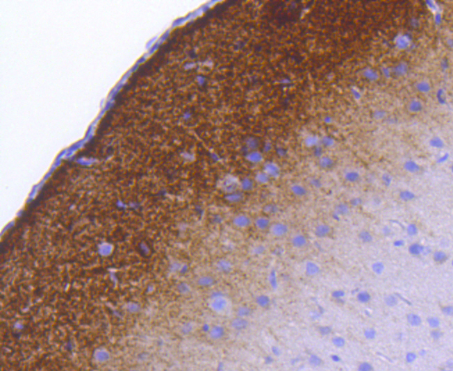
Immunohistochemical analysis of paraffin-embedded rat brain tissue using anti- phospho-Synapsin Ⅰ (S9) antibody. Counter stained with hematoxylin.IHC
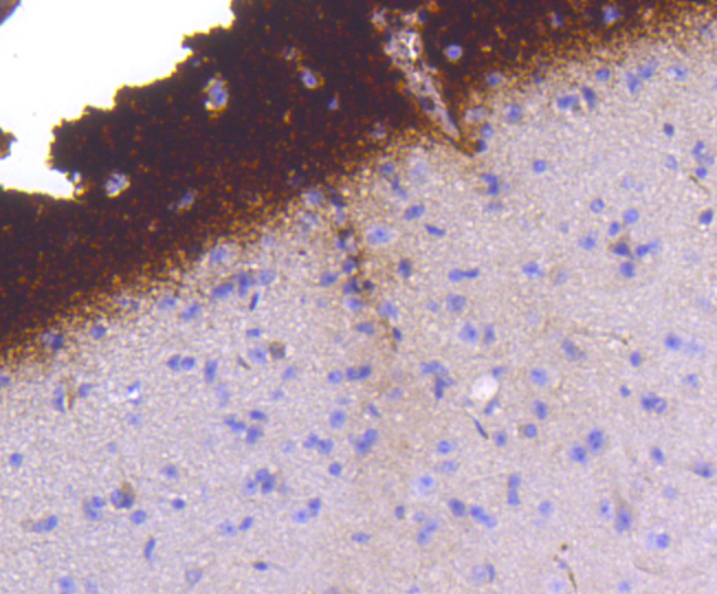
Immunohistochemical analysis of paraffin-embedded mouse brain tissue using anti- phospho-Synapsin Ⅰ (S9) antibody. Counter stained with hematoxylin.WB
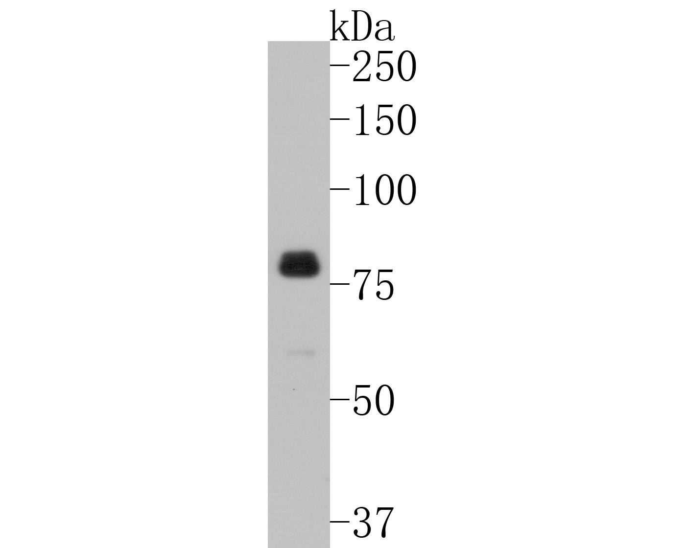
Western blot analysis of Phospho-Synapsin I (S9) on mouse brain tissue lysates with Rabbit anti-Phospho-Synapsin I (S9) antibody at 1/500 dilution. Lysates/proteins at 20 µg/Lane. Exposure time: 30 seconds; 8% SDS-PAGE gel. Proteins were transferred to a PVDF membrane and blocked with 5% NFDM/TBST for 1 hour at room temperature. The primary antibody at 1/500 dilution was used in 5% NFDM/TBST at room temperature for 2 hours. Goat Anti-Rabbit IgG - HRP Secondary Antibody at 1:200,000 dilution was used for 1 hour at room temperature.| Product Name | Phospho-Synapsin I (S9) Recombinant Rabbit Monoclonal Antibody |
|---|---|
| Antibody Type | Primary Antibodies |
| Product description | Synapsins are synaptic vesicle-associated phosphoproteins that regulate synaptic vesicle exocytosis and may be involved in synaptogenesis. Evidence suggests that Synapsin I, Synapsin II and Synapsin IIIa are ATP-binding proteins that are regulated by Ca2+ and calmodulin binding. Ca2+ has been shown to stimulate ATP binding to Synapsin I, to have no effect on Synapsin II and to inhibit Synapsin III. Synapsin I and Synapsin II both undergo alternative splicing to produce two forms of each protein, Synapsin la and lb and Synapsin IIa and IIb, respectively. Synapsin III gives rise to at least three isoforms: Synapsin IIIa, IIIb and IIIc. Synapsin III plays unique roles both in early axon outgrowth and in the regulation of synaptic vesicle trafficking. In cultured mouse hippocampal neurons, Synapsin III is expressed early during development, with levels peaking seven days after plating and declining thereafter. Synapsin III is highly concentrated in growth cones. |
| Immunogen | Synthetic phospho-peptide corresponding to residues surrounding Ser9 of Human Synapsin I aa 1-50 / 705. |
| Modification | p-S9 |
| Clonality | Monoclonal |
|---|---|
| Isotype | IgG |
| Host Species | Recombinant rabbit |
| Tested Applications | IHCWB |
| WB:1:500-1:1000 IHC:1:50-1:200 |
|
| Species Reactivity | HumanMouseRat |
| Concentration | 1mg/ml |
| Purification | Protein A |
| Alternative Names | Brain protein 4.1 antibody SYN 1 antibody SYN 1a antibody SYN 1b antibody SYN I antibody SYN1 antibody SYN1_HUMAN antibody SYN1a antibody SYN1b antibody Synapsin 1 antibody Synapsin I antibody Synapsin-1 antibody Synapsin1 antibody SynapsinI antibody SYNI antibody |
|---|---|
| Molecular Weight(MW) | 77 kDa |
| Cellular Localization | Cell junction, Golgi apparatus. |
IHC
Immunohistochemical analysis of paraffin-embedded rat brain tissue using anti- phospho-Synapsin Ⅰ (S9) antibody. Counter stained with hematoxylin.
IHC
Immunohistochemical analysis of paraffin-embedded mouse brain tissue using anti- phospho-Synapsin Ⅰ (S9) antibody. Counter stained with hematoxylin.
WB
Western blot analysis of Phospho-Synapsin I (S9) on mouse brain tissue lysates with Rabbit anti-Phospho-Synapsin I (S9) antibody at 1/500 dilution. Lysates/proteins at 20 µg/Lane. Exposure time: 30 seconds; 8% SDS-PAGE gel. Proteins were transferred to a PVDF membrane and blocked with 5% NFDM/TBST for 1 hour at room temperature. The primary antibody at 1/500 dilution was used in 5% NFDM/TBST at room temperature for 2 hours. Goat Anti-Rabbit IgG - HRP Secondary Antibody at 1:200,000 dilution was used for 1 hour at room temperature.| Positive Control | Rat brain tissue, mouse brain tissue. |
|---|---|
| Application Notes | WB:1:500-1:1000 IHC:1:50-1:200 |
| Form | Liquid |
|---|---|
| Storage Instructions | Store at +4℃ after thawing. Aliquot store at -20℃ or -80℃. Avoid repeated freeze / thaw cycles. |
| Storage Buffer | 1*TBS (pH7.4), 1%BSA, 40%Glycerol. Preservative: 0.05% Sodium Azide. |


