WB
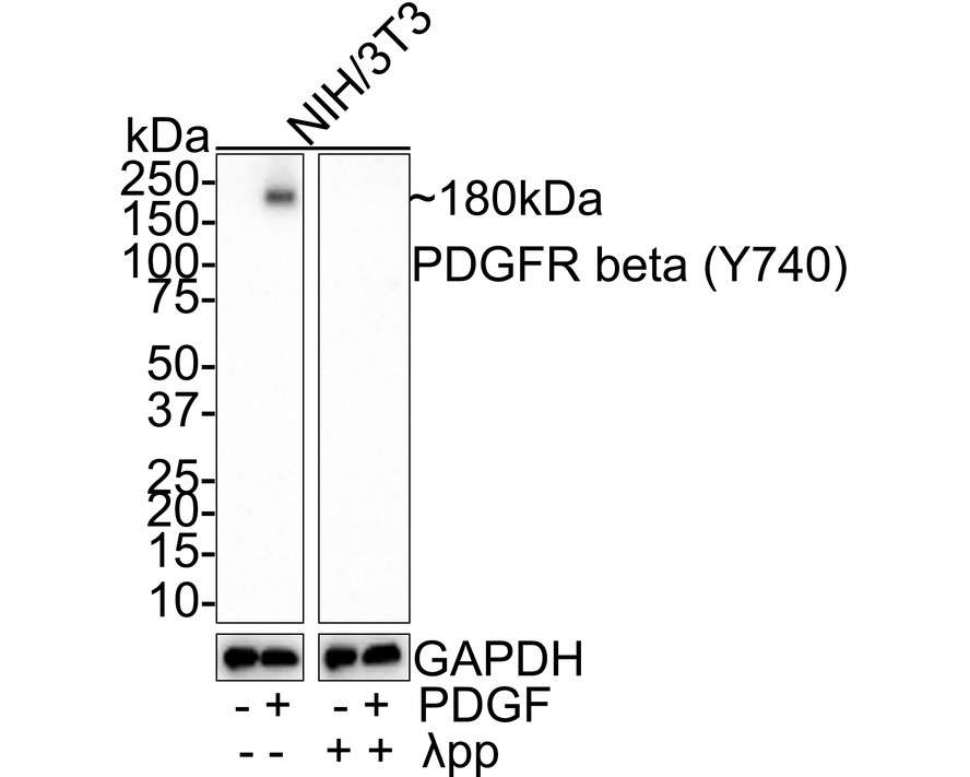
Western blot analysis of Phospho-PDGFR beta (Y740) on different lysates with Rabbit anti-Phospho-PDGFR beta (Y740) antibody at 1/1,000 dilution. Lane 1: NIH/3T3 whole cell lysate, Lane 2: NIH/3T3 treated with 50 ng/mL PDGF for 5 minutes whole cell lysate, Lane 3: NIH/3T3 treated with λpp whole cell lysate, Lane 4: NIH/3T3 treated with 50 ng/mL PDGF for 5 minutes then treated with λpp whole cell lysate, Lysates/proteins at 20 µg/Lane. Exposure time: 2 minutes; 4-20% SDS-PAGE gel. Proteins were transferred to a PVDF membrane and blocked with 5% NFDM/TBST for 1 hour at room temperature. The primary antibody at 1/1,000 dilution was used in 5% NFDM/TBST at room temperature for 2 hours. Goat Anti-Rabbit IgG - HRP Secondary Antibody at 1:100,000 dilution was used for 1 hour at room temperature.ICC/IF
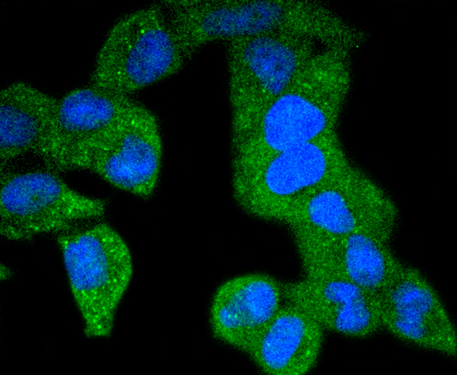
ICC staining PDGF Receptor beta(phospho Y740) in Hela cells (green). The nuclear counter stain is DAPI (blue). Cells were fixed in paraformaldehyde, permeabilised with 0.25% Triton X100/PBS.ICC/IF
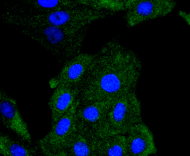
ICC staining PDGF Receptor beta(phospho Y740) in L6 cells (green). The nuclear counter stain is DAPI (blue). Cells were fixed in paraformaldehyde, permeabilised with 0.25% Triton X100/PBS.| Product Name | Phospho-PDGFR beta (Y740) Recombinant Rabbit Monoclonal Antibody |
|---|---|
| Antibody Type | Primary Antibodies |
| Product description | Platelet Derived Growth Factor (PDGF) is a mitogen for mesenchyme- and glia-derived cells. PDGF consists of two chains, A and B, which dimerize to form functionally distinct isoforms, PDGF-AA, PDGF-AB, and PDGF-BB. These three isoforms bind with different affinities to two receptor types, a and b, which are endowed with protein tyrosine kinase domains and undergo either homo- or heterodimerization as a consequence of ligand binding. Ligand stimulation of PDGFR-b leads to autophosphorylation at Tyr-857, which is the major autophosphorylation site, and Tyr-751, which is the major in vitro phosphorylation site. Autophosphorylation of Tyr-751, which lies in the kinase insert region, is required for binding of phosphatidylinositol-3 kinase to the receptor. These auto-phosphorylation events largely contribute to signal transduction through the PDGF receptor. |
| Immunogen | Synthetic phospho-peptide corresponding to residues surrounding Tyr740 of human PDGFR beta. |
| Modification | p-Y740 |
| Clonality | Monoclonal |
|---|---|
| Isotype | IgG |
| Host Species | Recombinant rabbit |
| Tested Applications | ICC/IFWB |
| WB:1:1000 ICC/IF:1:50-1:200 |
|
| Species Reactivity | HumanMouseRat |
| Concentration | 1mg/ml |
| Purification | Protein A |
| Alternative Names | Beta platelet derived growth factor receptor antibody Beta-type platelet-derived growth factor receptor antibody CD 140B antibody CD140 antigen-like family member B antibody CD140b antibody CD140b antigen antibody IBGC4 antibody IMF1 antibody JTK12 antibody OTTHUMP00000160528 antibody PDGF R beta antibody PDGF-R-beta antibody PDGFR 1 antibody PDGFR antibody PDGFR beta antibody PDGFR1 antibody PDGFRB antibody PGFRB_HUMAN antibody Platelet derived growth factor receptor 1 antibody Platelet derived growth factor receptor beta antibody Platelet derived growth factor receptor beta polypeptide antibody |
|---|---|
| Molecular Weight(MW) | 120 kDa(Observed band size: 180 kDa) |
| Cellular Localization | Cell membrane, Cytoplasmic vesicle. |
WB
Western blot analysis of Phospho-PDGFR beta (Y740) on different lysates with Rabbit anti-Phospho-PDGFR beta (Y740) antibody at 1/1,000 dilution. Lane 1: NIH/3T3 whole cell lysate, Lane 2: NIH/3T3 treated with 50 ng/mL PDGF for 5 minutes whole cell lysate, Lane 3: NIH/3T3 treated with λpp whole cell lysate, Lane 4: NIH/3T3 treated with 50 ng/mL PDGF for 5 minutes then treated with λpp whole cell lysate, Lysates/proteins at 20 µg/Lane. Exposure time: 2 minutes; 4-20% SDS-PAGE gel. Proteins were transferred to a PVDF membrane and blocked with 5% NFDM/TBST for 1 hour at room temperature. The primary antibody at 1/1,000 dilution was used in 5% NFDM/TBST at room temperature for 2 hours. Goat Anti-Rabbit IgG - HRP Secondary Antibody at 1:100,000 dilution was used for 1 hour at room temperature.
ICC/IF
ICC staining PDGF Receptor beta(phospho Y740) in Hela cells (green). The nuclear counter stain is DAPI (blue). Cells were fixed in paraformaldehyde, permeabilised with 0.25% Triton X100/PBS.
ICC/IF
ICC staining PDGF Receptor beta(phospho Y740) in L6 cells (green). The nuclear counter stain is DAPI (blue). Cells were fixed in paraformaldehyde, permeabilised with 0.25% Triton X100/PBS.| Application Notes | WB:1:1000 ICC/IF:1:50-1:200 |
|---|
| Form | Liquid |
|---|---|
| Storage Instructions | Store at +4℃ after thawing. Aliquot store at -20℃ or -80℃. Avoid repeated freeze / thaw cycles. |
| Storage Buffer | 1*TBS (pH7.4), 1%BSA, 40%Glycerol. Preservative: 0.05% Sodium Azide. |


