Application
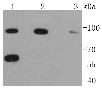
Fig1: Western blot analysis of RAD18 on different lysates using anti-RAD18 antibody at 1/1,000 dilution. Positive control: Lane 1: NIH/3T3 Lane 2: 293T Lane 3: MCF-7Application
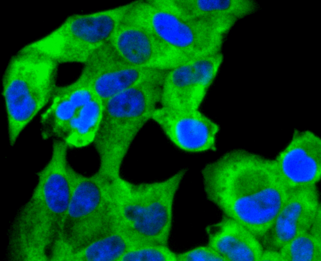
Fig2: ICC staining RAD18 in Hela cells (green). The nuclear counter stain is DAPI (blue). Cells were fixed in paraformaldehyde, permeabilised with 0.25% Triton X100/PBS.Application
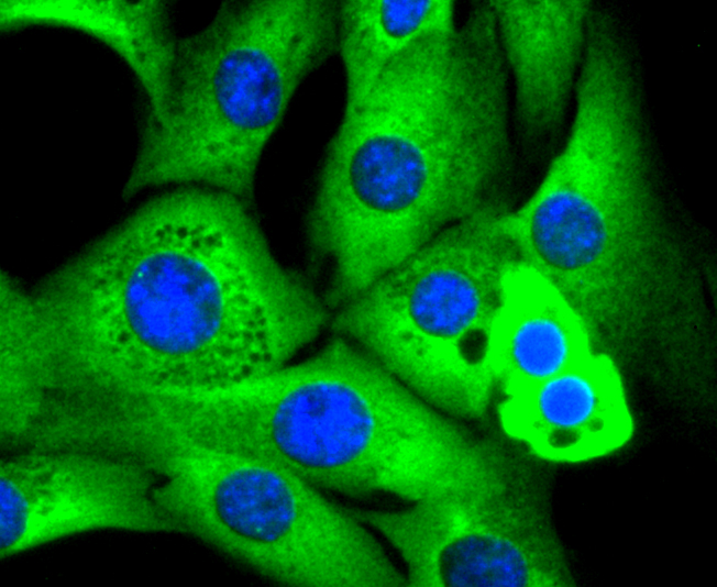
Fig3: ICC staining RAD18 in NIH/3T3 cells (green). The nuclear counter stain is DAPI (blue). Cells were fixed in paraformaldehyde, permeabilised with 0.25% Triton X100/PBS.Application
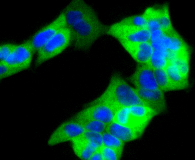
Fig4: ICC staining RAD18 in 293 cells (green). The nuclear counter stain is DAPI (blue). Cells were fixed in paraformaldehyde, permeabilised with 0.25% Triton X100/PBS.Application
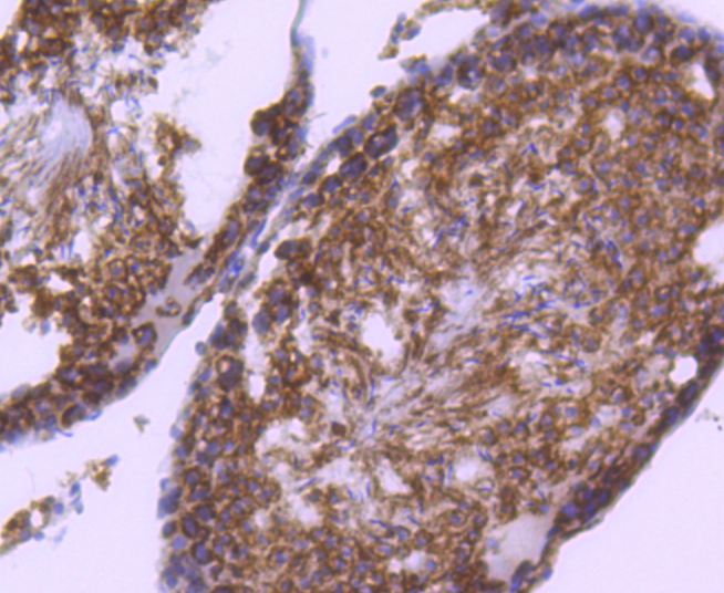
Fig5: Immunohistochemical analysis of paraffin-embedded rat testis tissue using anti-RAD18 antibody. Counter stained with hematoxylin.Application
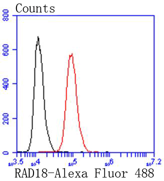
Fig6: Flow cytometric analysis of Hela cells with RAD18 antibody at 1/50 dilution (red) compared with an unlabelled control (cells without incubation with primary antibody; black). Alexa Fluor 488-conjugated goat anti rabbit IgG was used as the secondary antibody| Product Name | RAD18 [SN66-01] |
|---|---|
| Antibody Type | Primary Antibodies |
| Antigen Alias | 2810024C04Rik antibody DNA repair protein rad18 antibody E3 ubiquitin-protein ligase RAD18 antibody EC 6.3.2.- antibody hHR 18 antibody hHR18 antibody hRAD 18 antibody hRAD18 antibody MGC156682 antibody Postreplication repair E3 ubiquitin-protein ligase RAD18 antibody Postreplication repair protein hRAD18p antibody Postreplication repair protein RAD18 antibody RAD 18 antibody RAD18 antibody RAD18 homolog (S. cerevisiae) antibody RAD18 homolog antibody RAD18 S. cerevisiae homolog antibody RAD18 S. cerevisiae homolog of antibody RAD18 transcript variant antibody RAD18_HUMAN antibody Rad18sc antibody Radiation sensitivity protein 18 antibody RING finger protein 73 antibody RNF 73 antibody RNF73 antibody Structural maintenance of chromosomes protein 6 antibody YCR066W antibody YCR66W antibody |
| Product description | The RING-type zinc finger protein Rad18 is essential in post-replication repair of damaged DNA and contributes to the maintenance of genomic stability. Rad18 maintains chromosomal DNA with the Rad54-dependent DNA repair pathway and recruits ubiquitin-conjugating enzymes in the Rad6 pathway. Rad18 functions in gap-filling of a daughter strand on replication of damaged DNA and is localized in the nucleus. |
| Immunogen | recombinant protein |
| Clonality | Monoclonal |
|---|---|
| Isotype | IgG |
| Host Species | Recombinant rabbit |
| Tested Applications | WBICC/IFIHCFC |
| WB:1:1,000-1:2,000 ICC:1:100-1:500 IHC:1:50-1:200 FC:1:50-1:100 |
|
| Species Reactivity | HumanMouseRat |
| Concentration | 1mg/ml |
| Alternative Names | 2810024C04Rik antibody DNA repair protein rad18 antibody E3 ubiquitin-protein ligase RAD18 antibody EC 6.3.2.- antibody hHR 18 antibody hHR18 antibody hRAD 18 antibody hRAD18 antibody MGC156682 antibody Postreplication repair E3 ubiquitin-protein ligase RAD18 antibody Postreplication repair protein hRAD18p antibody Postreplication repair protein RAD18 antibody RAD 18 antibody RAD18 antibody RAD18 homolog (S. cerevisiae) antibody RAD18 homolog antibody RAD18 S. cerevisiae homolog antibody RAD18 S. cerevisiae homolog of antibody RAD18 transcript variant antibody RAD18_HUMAN antibody Rad18sc antibody Radiation sensitivity protein 18 antibody RING finger protein 73 antibody RNF 73 antibody RNF73 antibody Structural maintenance of chromosomes protein 6 antibody YCR066W antibody YCR66W antibody |
|---|---|
| Molecular Weight(MW) | 56/90kDa |
| Cellular Localization | Nucleus, Cytoplasm. |
Application
Fig1: Western blot analysis of RAD18 on different lysates using anti-RAD18 antibody at 1/1,000 dilution. Positive control: Lane 1: NIH/3T3 Lane 2: 293T Lane 3: MCF-7
Application
Fig2: ICC staining RAD18 in Hela cells (green). The nuclear counter stain is DAPI (blue). Cells were fixed in paraformaldehyde, permeabilised with 0.25% Triton X100/PBS.
Application
Fig3: ICC staining RAD18 in NIH/3T3 cells (green). The nuclear counter stain is DAPI (blue). Cells were fixed in paraformaldehyde, permeabilised with 0.25% Triton X100/PBS.
Application
Fig4: ICC staining RAD18 in 293 cells (green). The nuclear counter stain is DAPI (blue). Cells were fixed in paraformaldehyde, permeabilised with 0.25% Triton X100/PBS.
Application
Fig5: Immunohistochemical analysis of paraffin-embedded rat testis tissue using anti-RAD18 antibody. Counter stained with hematoxylin.
Application
Fig6: Flow cytometric analysis of Hela cells with RAD18 antibody at 1/50 dilution (red) compared with an unlabelled control (cells without incubation with primary antibody; black). Alexa Fluor 488-conjugated goat anti rabbit IgG was used as the secondary antibody| Positive Control | NIH/3T3, MCF-7, 293T. |
|---|---|
| Application Notes | WB:1:1,000-1:2,000 ICC:1:100-1:500 IHC:1:50-1:200 FC:1:50-1:100 |
| Form | Liquid |
|---|---|
| Storage Instructions | Store at +4℃ after thawing. Aliquot store at -20℃ or -80℃. Avoid repeated freeze / thaw cycles. |
| Storage Buffer | 1*TBS (pH7.4), 1%BSA, 40%Glycerol. Preservative: 0.05% Sodium Azide. |


