Application
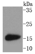
Fig1: Western blot analysis of MAP1LC3A on mouse brain lysates using anti-MAP1LC3A antibody at 1/1,000 dilution.Application
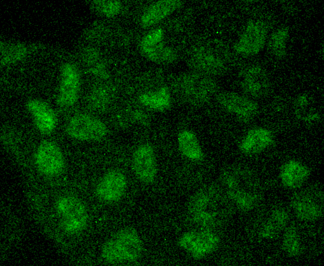
Fig2: ICC staining MAP1LC3A in Hela cells (green). The nuclear counter stain is DAPI (blue). Cells were fixed in paraformaldehyde, permeabilised with 0.25% Triton X100/PBS.Application
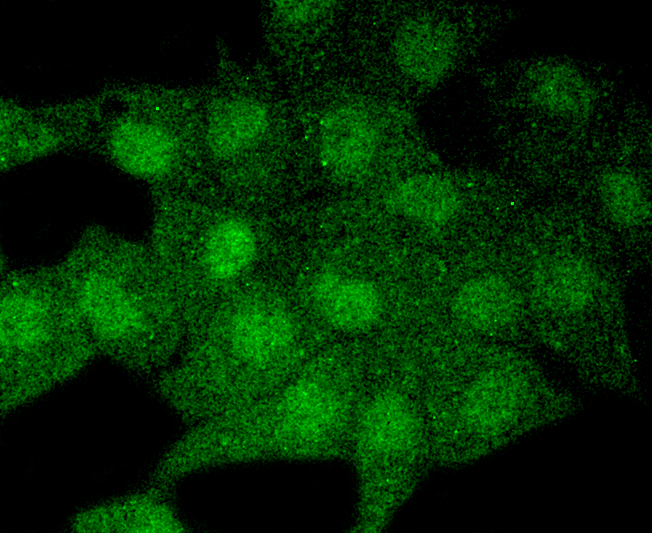
Fig3: ICC staining MAP1LC3A in PMVEC cells (green). The nuclear counter stain is DAPI (blue). Cells were fixed in paraformaldehyde, permeabilised with 0.25% Triton X100/PBS.Application
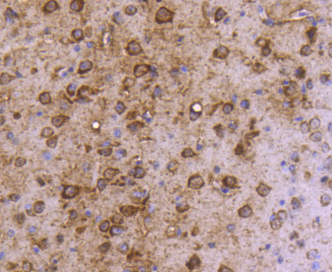
Fig4: Immunohistochemical analysis of paraffin-embedded mouse brain tissue using anti-MAP1LC3A antibody. Counter stained with hematoxylin.| Product Name | MAP1LC3A [SN07-40] |
|---|---|
| Antibody Type | Primary Antibodies |
| Antigen Alias | ATG8E antibody Autophagy-related protein LC3 A antibody Autophagy-related ubiquitin-like modifier LC3 A antibody LC3 antibody LC3A antibody MAP1 light chain 3 like protein 1 antibody MAP1 light chain 3-like protein 1 antibody MAP1A/1B light chain 3 A antibody MAP1A/MAP1B LC3 A antibody MAP1A/MAP1B light chain 3 A antibody MAP1ALC3 antibody MAP1BLC3 antibody Map1lc3a antibody Microtubule associated proteins 1A/1B light chain 3 antibody Microtubule-associated protein 1 light chain 3 alpha antibody Microtubule-associated proteins 1A and 1B, light chain 3 antibody Microtubule-associated proteins 1A/1B light chain 3A antibody MLP3A_HUMAN antibody |
| Product description | Microtubule-associated proteins (MAPs) regulate microtubule stability and play critical roles in neuronal development and in maintaining the balance between neuronal plasticity and rigidity. MAP-light chain 3 beta (MAP-LC3β) and MAP-light chain 3 alpha (MAP-LC3α), both of which are mammalian homologs of yeast Apg8, are subunits that can associate with either MAP-1A or MAP-1B. While MAP-LC3β is essential for autophagy and is associated with autophagosome membranes after processing, MAP LC3α is involved in the formation of autophagosomal vacuoles and is localized to the intracytoplasmic membrane. MAP LC3α is expressed as two alternatively spliced isoforms that are expressed in testis, brain, heart, liver and skeletal muscle, but are absent in thymus and peripheral blood leukocytes. |
| Immunogen | recombinant protein |
| Clonality | Monoclonal |
|---|---|
| Isotype | IgG |
| Host Species | Recombinant rabbit |
| Tested Applications | WBICCIHCIP |
| WB:1:1,000-1:2,000 ICC:1:50-1:200 IHC:1:50-1:200 |
|
| Species Reactivity | HumanMouseRat |
| Concentration | 1mg/ml |
| Alternative Names | ATG8E antibody
Autophagy-related protein LC3 A antibody
Autophagy-related ubiquitin-like modifier LC3 A antibody
LC3 antibody
LC3A antibody
MAP1 light chain 3 like protein 1 antibody
MAP1 light chain 3-like protein 1 antibody
MAP1A/1B light chain 3 A antibody
MAP1A/MAP1B LC3 A antibody
MAP1A/MAP1B light chain 3 A antibody
MAP1ALC3 antibody
MAP1BLC3 antibody
Map1lc3a antibody
Microtubule associated proteins 1A/1B light chain 3 antibody
Microtubule-associated protein 1 light chain 3 alpha antibody
Microtubule-associated proteins 1A and 1B light chain 3 antibody Microtubule-associated proteins 1A/1B light chain 3A antibody MLP3A_HUMAN antibody |
|---|---|
| Molecular Weight(MW) | 14 kDa |
| Cellular Localization | Cytoplasm, Endomembrane system, Cytoplasmic vesicle. |
Application
Fig1: Western blot analysis of MAP1LC3A on mouse brain lysates using anti-MAP1LC3A antibody at 1/1,000 dilution.
Application
Fig2: ICC staining MAP1LC3A in Hela cells (green). The nuclear counter stain is DAPI (blue). Cells were fixed in paraformaldehyde, permeabilised with 0.25% Triton X100/PBS.
Application
Fig3: ICC staining MAP1LC3A in PMVEC cells (green). The nuclear counter stain is DAPI (blue). Cells were fixed in paraformaldehyde, permeabilised with 0.25% Triton X100/PBS.
Application
Fig4: Immunohistochemical analysis of paraffin-embedded mouse brain tissue using anti-MAP1LC3A antibody. Counter stained with hematoxylin.| Positive Control | PMVEC, Hela, mouse brain tissue. |
|---|---|
| Application Notes | WB:1:1,000-1:2,000 ICC:1:50-1:200 IHC:1:50-1:200 |
| Form | Liquid |
|---|---|
| Storage Instructions | Store at +4℃ after thawing. Aliquot store at -20℃ or -80℃. Avoid repeated freeze / thaw cycles. |
| Storage Buffer | 1*TBS (pH7.4), 1%BSA, 40%Glycerol. Preservative: 0.05% Sodium Azide. |


